แสดงบทความที่มีป้ายกำกับ tennis แสดงบทความทั้งหมด

วันพุธที่ 8 มิถุนายน พ.ศ. 2565

Physiotherapy with 9 options to stretch quadriceps femoris


Quadriceps Femoris strain in soccer players
Ref: https://talksport.com/football/

Quadriceps Femoris (QF) composes the main part of the anterior muscles of the thigh, being a powerful extensor of the leg, considered the most beautiful and largest and most powerful muscle. Its name is derived from Latin, hip  +  caput, or thigh muscle with four heads. 

The quadriceps femoris (QF) is a major extensor of the knee joint that is crucial for human movements. There are sharing working functions in the locomotion, for example, the rectus femoris (RF) swings the leg forward when a step is taken. During walking or running, quadriceps muscles such as the vastus medialis (VM) stabilize the patella and knee joint.  

Quadriceps Femoris anatomy
(Ref: https://www.corewalking.com/rectus-femoris-tendon/)


“Its main functions include knee extension and hip flexion”. 


The QF is important in sport owing to its potential for injury, which can be painful and debilitating. Strains, tears, and contusions are relatively common and require recovery time. I underwent treatment caring for professional soccer players with proximal and distal QF strain or inflammation. In proximal injury cases, I have seen muscle strain at the origin of the tendon of QF. In distal injury cases, I have seen patellar tendonitis and patellofemoral pain. I rarely have seen muscle strain at mid QF muscle in soccer players, but opposite in sprint runners. 

Ball shooting composes hip flexion and knee extension
(Ref: https://scialert.net/fulltext/?doi=jas.2010.1286.1292)


I have used stretching not only physiotherapy treatment and prevention for QF but also hamstring strain prevention. Moreover, it has been applied to my many case e.g. IT band syndrome and low back pain.


9 options to stretch quadriceps femoris


Exercise #1: Half kneeling QF stretching; target leg is rear that need some cushion under it. For more comfortable, put opposite hand on any stable furnitures or wall for balance. During stretching, try to set hip joint to be neutral or extension.


Exercise #2: Stand QF stretch: For more comfortable, put opposite hand on any stable furnitures or wall for balance. During stretching, try to set hip joint to be neutral or extension.


Exercise #3: Chair single kneeling QF stretch: During stretching, try to set hip joint to be neutral or extension.


Exercise #4: Supine with double knees bending QF stretch: You can try to lying on the back if possible.


Exercise #5: Supine with double knees bending QF stretch: You can try to lying on the back if possible.


Exercise #6: Side lying QF stretch: the target leg is above. During stretching, try to set hip joint to be neutral or extension.


Exercise #7: Prone QF stretch


Exercise #8: Side lying QF stretch with cable assisted: During stretching, try to set hip joint to be neutral or extension.


Exercise #9: Prone QF stretch with cable assisted: During stretching, try to set hip joint to be neutral or extension.


The members of quadriceps femoris consists of one rectus plus three vasti that are sum of four; include,


(1) Rectus Femoris (RF)

The RF is a long, fusiform muscle forming the anterior superficial portion of the quadriceps muscle group that originates from the small area of bony pelvis which is the anterior inferior iliac spine (AIIS). The proximal rectus femoris has two tendinous origins: the direct (straight) head, arising from the anterior–inferior iliac spine, and the indirect (reflected) head, arising slightly more inferiorly and posteriorly from the superior acetabular ridge and hip joint capsule.

The Superficial Layer of the Quadriceps Tendon The RF, the most superficial muscle of the quadriceps group, inserts into the anterior portion of the base and the superior third of the anterior surface of the patella

Due to the RF being the most superficial muscle of the QF complex, its insertion is on a layer of the superficial layer of the quadriceps tendon that is attached into the anterior portion of the base and the superior third of the anterior surface of the patella.

Its main functions include knee extension and hip flexion. 

Rectus Femoris muscle anatomy
(Ref: https://pixels.com/)


(2) Vastus Intermedius (VI)

VI typically arose muscularly from the anterior and lateral aspect of the femur without an origin tendon. The VI originates from the upper two-thirds of the anterior and lateral surfaces of the femur and the intermuscular septum and forms the quadriceps tendon. The shape and area of the origin domain of the VI was much more diverse than those of the vastus lateralis and vastus medialis.

Classification of the origin domain of the VI into contacting and non-contacting types on the basis of its positional relationship to the origin domain of vastus lateralis. The origin domain of the contact type attached muscularly to the anterior and lateral surface of the femoral shaft. The muscle belly decreased in thickness from its anterior to its lateral parts. The origin domain of the VI adjoined the longus part of vastus lateralis at the lateral line of linea aspera to form a common origin domain. The muscle belly and origin domain of the non - contacting type were much smaller than the contacting type; they were narrow and almost corresponded to the width of the femoral shaft. The origin domain of the VI attached only to the anterior surface of the femur and did not contact the lateral line of the linea aspera and lay between the origin domains of the VI and VL.

Vastus Intermedius muscle anatomy
(Ref: https://www.getbodysmart.com/leg-muscles/vastus-intermedius-muscle)


Insertion of VI involves insertion of VL, VM, and RF that are located in the deep layer of the quadriceps tendon. The VI has an intimate origin with VL proximally and the lateral intermuscular septum distally. It inserts through a broad, thin tendon into the base of the patella posterior to the VL and VM or merged with them. The VI also may join with the RF to form the suprapatellar tendon, which inserts on the base of the patella. Medially and laterally, this insertion reinforces the patellofemoral ligaments.


(3) Vastus Medialis (VM)

The VM seems to be a complicated muscle which stays at the anterior surface of the femur. It originates on the lower part of the intertrochanteric line, and the upper third of the medial supracondylar line and has attachments to the medial lip of the linea aspera as it wraps around the femur. 

The division of the vastus in two parts, one long and one oblique. VM was identified into the VML and VMO by three features identification; include, fiber angle, the presence or absence of a fascial plane of separation, and the pattern of innervation. The trajectory of the muscle fibers of the vastus medialis split into  3 parts: the first would be  1/3 of the top is composed of parallel fibers that arise vertically from the intertrochanteric line, converging toward the top edge of a muscle aponeurosis found deeply, the second would be  1/3 middle part which is composed of parallel fibers obliquely, running about 15-35° to the longitudinal axis of the femur, arising from the lip of the linea aspera femoris and the medial intermuscular septum inserting the medial margin and the anterior surface of the aponeurosis found and, finally, the third, which would be the bottom 1/3 of the muscle was composed of parallel fibers being in mostly aligned more obliquely to 40-55° to the longitudinal axis of the femur presenting appearance almost horizontal, arising mainly from the adductor magnus tendon and partially on the medial intermuscular septum. The vastus medialis oblique is the distal part having its origin mainly in the adductor magnus tendon and its insertion into the medial margin of the patella.

Vastus Medialis Longus is medial to Vastus Medialis Oblique
(Ref: https://alliedanatomy.com/)


Insertion of VM is in the intermediate layer of the quadriceps tendon. The most inserted fibers of the VM end in an aponeurosis that blends with the medial side of the suprapatellar tendon or the RF tendon. They attach directly to the medial edge of the patella and extend more distally than fibers originating from any other part of the quadriceps group. From the distal edge of the VM, a tendinous expansion passes along the medial side of the patella. Deep fibers of this expansion reinforce the joint capsule as part of the medial patellar retinaculum. 

Vastus medialis longus is responsible for knee extension, whereas the primary function of the vastus medialis oblique (VMO) is medial stabilization of the patellar throughout extension. Medial stabilization is the function of preventing a lateral subluxation of the patella by maintaining the alignment of the patella to slip on the femoral condyles, providing medial force directly to counterbalance the forces directed laterally by the vastus lateralis. Therefore, VMO is considered  an acceptable solution to problems involving the rehabilitation of the stabilization of the patella. 


(4) Vastus Lateralis (VL). 

VL is considered the largest part of the quadriceps femoris to form thick, wide, and fusiform appearance is intimately attached to the vastus intermedius in its middle third.

The origin of the vastus lateralis is on the greater trochanter, lateral lip of linea aspera femoris and the lateral intermuscular septum and its insertion at the base patellar ligament using the patellar and tibial tuberosity.

Vastus lateralis muscle has presented two parts; include, long and oblique parts. The oblique portion arises from the linea aspera of the femur and lateral intermuscular septum, a more fibrous expansion of the iliotibial tract by insertion the tendon itself, which is directed inferior and lateral to the VL over this by joining the superolateral border of the patellar tendon to form a single, often attached to the joint capsule. The distal fibers of the vastus lateralis oblique Were presented in interdigitated lateral retinaculum, which is formed from the expansions of tedious insertions of the VL, inserted in the tibia and patella which, in turn, reinforces the anterolateral region of knee, and iliotibial tract. Morphologically, the vastus lateralis oblique is divided into: a distal surface and a spiral proximal portion with deep longitudinal fibers in the femoral shift below the belly of the VL along, mingling with the vastus intermedius.

Vastus Lateralis muscle anatomy
(Ref: https://learnmuscles.com/glossary/vastus-lateralis/)


The vastus lateralis longus has its origin in the femoral trochanter and lateral lip of linea aspera of the femur, as found in the anatomical literature showing extensive superficial aponeurosis in more than two thirds of the proximal surface and a deep aponeurosis in the distal one thirds separating it from vastus lateralis oblique. Its insertion is on the superolateral border of the patellar ligament by means of union with the tendon of vastus lateralis oblique, forming a single tendon.

The VL muscle consists of three layers: superficial, intermediate, and deep: (i) The superficial part originates from the lateral surface of the greater trochanter. Its fibers arch medially downward to the tendon lamina and then to the QF tendon (ii) The intermediate part originates from the upper level of the greater trochanter’s anterior surface where it joins the intertrochanteric and gluteus medius ridge (iii) The deep part is the one-third proximal to the femur.

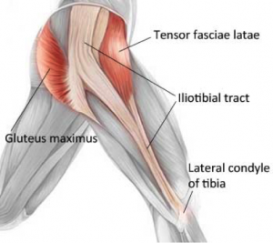
Vastus lateralis beneath IT band
(Ref: https://www.shutterstock.com/)


The distal insertion of the VL and VM unite to form a continuous aponeurosis that inserts into the base of the patella, just posterior to the insertion of RF, and also continues laterally and medially to insert into the sides of the patella that happen in the iIntermediate layer of the quadriceps tendon. The VL ends in an aponeurosis that blends with the lateral side of the suprapatellar or RF tendon and sends an expansion distally to the superolateral side of the patella. Other fibers cross superficial to the patella and attach to the medial condyle of the tibia. The lateral expansion of VL then blends with the capsule of the knee, thereby forming part of the lateral patellar retinaculum. 


Conclusion, RF is two joint muscles of hip and knee that responsible for hip flexion with knee extension. Others are one joint muscle of knee that function for knee extension.


In my physiotherapy experience, stretching this muscle was not as easy as many muscles because it did not provide obvious tension. However, the principle to stretch this muscle is the same as the others: stretch to the point where “tightness with pain” or “noticeable tension without pain” will hole at the point for 30 seconds of 3 - 5 reputations as demonstrated VIDEO.


Reference:

https://www.researchgate.net/publication/307885598_Structural_diversity_of_the_vastus_intermedius_origin_revealed_by_analysis_of_isolated_muscle_specimens 

https://www.ncbi.nlm.nih.gov/pmc/articles/PMC2941577/ 

https://www.ncbi.nlm.nih.gov/pmc/articles/PMC8404765/ 

https://www.hindawi.com/journals/bmri/2022/9569101/ 

https://www.ncbi.nlm.nih.gov/pmc/articles/PMC2772911/ 

https://www.scielo.br/j/rbme/a/htP9y5rrfQdkGFLDkZPQF6r/?lang=en&format=pdf

http://www.jms.periodikos.com.br/article/587cb49f7f8c9d0d058b47a1/pdf/jms-28-4-587cb49f7f8c9d0d058b47a1.pdf 

https://www.ajronline.org/doi/pdf/10.2214/AJR.07.2947 

https://www.researchgate.net/publication/234012902_Vastus_medialis_a_reappraisal_of_VMO_and_VML 


วันศุกร์ที่ 27 พฤษภาคม พ.ศ. 2565

To release IT band syndrome by 5 ways foam roll massage


Ref: https://www.runwell.net.au/blog/

        Foam roll massage and tennis ball massage is a popular technique of muscle - fascia tension release for prevent and treatment benefits. It is applied to many parts of the body and musculoskeletal impairments including the IT band syndrome. 

In my physiotherapy clinic experience with IT band syndrome patients, I have used this technique many times that roll on the IT band directly as the standard management. The outcomes demonstrated both effectiveness and ineffectiveness. Finally, I found a way to massage with the foam that I always recommend to patients who are with IT band syndrome. 

Ref: https://physioworks.com.au/pain-injury/knee-pain/itb-friction-syndrome/


I have seen muscle spasm not only at mid IT band but also above knee cap, rear hip joint, calf muscles. 

5 ways to do roll massage in IT band syndrome

Exercise #1: IT band: lying on the side to roll.


Exercise #2: Gluteus maximus: it is necessary to bend hip joint with half - prone in front.


Exercise #3: Tensor Fascia Lata: prone on the ball to roll.


Exercise #4: Biceps femoris: lying on the side with half - supine.


Exercise #5: Vastus lateralis: lying on the side with half - prone.


I would like to introduce all muscles anatomy  which I have recommended to do foam roll; include,

    (1) The IT band

    IT band is a strong thick wide tendinous band located on the lateral thigh. IT band origin is insertion attachment of gluteus maximus (Gmax) and Tensor Fascia Latae (TFL) that extend their part into the IT band directly. The mass of the IT band descended along the lateral thigh to the knee joint that covers the vastus lateralis muscle. The IT band has 2 primary attachments, including the lateral epicondyle and the Gerdy tubercle. 

Ref: https://www.mygcphysio.com.au/


The first iliotibial band attachment is into the distal femur at the upper edge of the lateral epicondyle. The histologic makeup is consistent with tendon and has a layer of adipose tissue underneath the iliotibial band attachment area. The adipose tissue contains pacinian corpuscles, is highly vascular, and may be “the site of the inflammation” that causes pain during compression.

The second attachment of the iliotibial band is the insertion into the Gerdy tubercle of the tibia and serves as a ligament in structure and function. The Gerdy tubercle attachment is tensed during tibia internal rotation as the knee flexes during the weight-acceptance phase of gait. Internal tibial rotation explains the occasional connection between toeing in and iliotibial band “strain”

Moreover, there are many distal attachments that span out to the lateral border of the patella (by way of epicondylopatellar ligament and patellar retinaculum), patellar tendon, lateral patellar retinaculum before its insertion on Gerdy’s tubercle of the tibia, the biceps femoris, and vastus lateralis. 

About this muscle, I have seen trigger points at mid IT band and 2 - 5 cm. above knee joint the most. 


(2) Gluteus maximus (Gmax)

We can see Gmax at the rear hip which is the biggest muscle that is why origin attachment of Gmax covered large area; include, posterior ilium, posterior layer of thoracolumbar fascia, posterior superior iliac spine (PSIS), posterior gluteal line, posteroinferior sacrum, coccyx, sacrotuberous ligament, and over on gluteal fascia. It is used for support and bumpers while sitting in our daily lives. Gmax has two distinct portions that consist of inserts into the IT band and the inferior portion inserts into the femur. The Gmax pulls posteriorly on the IT band and femur to extend the hip.    

According to its insertion, there is thickening and stiff soft tissue behind the greater trochanter. 


     (3)Tensor Fascia Lata (TFL)

Origin of TFL started on anterolateral of an iliac crest that is approximately 15 - 76 millimeters wide. TFL has many origin attachments; include, the lateral aspect of the anterior superior iliac spine (ASIS). The iliac tubercle, a notch located below the superior spine of the ilium, the anterolateral iliac fossa that is slightly below the crest, and the deep surface of the fascia lata of the thigh. 

Insertion of TFL has been recorded in many locations. There are disputes , for example, on greater trochanter or around there, to the middle layer of the IT band. So, It is not clear whether such tendinous tissue belongs to the fascia lata, or is the muscle’s own tendon. However, the TFL pulls anterosuperiorly on the ITB to flex the hip generally.

I have found tightness, stiffness, and trigger points on this muscle often that related IT band tension progressively.  

Ref: https://www.pinterest.com/pin/


(4) Biceps femoris

Biceps femoris is a member of the hamstring muscle which is located on the lateral side of the posterior thigh. They consist of long head and short head that short head originated medial to the linea aspera of the distal femur and descended distally and laterally. 

The first component of the short head was a proximal muscular attachment to the anterior and medial side of the tendon of the long head. Other significant insertions included an attachment of the capsular arm to the posterolateral joint capsule,  the attachment of the capsuloosseous layer to the iliotibial tract (biceps-capsuloosseous iliotibial tract confluens),  a lateral aponeurosis, two tendinous attachments-the direct arm,  the anterior arm, and capsular attachment in the interval between the tendon of the lateral head of the gastrocnemius. Perhaps, insertion anatomy caused patients with IT band syndrome to feel tense calf muscle. 

My own opinion, it is not clear between biceps femoris tightness or posterior fiber of IT band stiffness. 

Ref: https://anatomy.app/encyclopedia/biceps-femoris


(5) Vastus lateralis (VL)

VL is a member of quadriceps composed structurally of four distinct muscles; include, the rectus femoris, vastus lateralis, vastus medialis and vastus intermedius. The vastus lateralis is considered the largest part of the quadriceps femoris to form thick, wide and fusiform appearance is intimately attached to the vastus intermedius in its middle third. 

          The origin of the vastus lateralis is on the greater trochanter, lateral lip of linea aspera femoris and the lateral intermuscular septum and its insertion at the base patellar ligament using the patella and tibial tuberosity.

Vastus lateralis muscle has presented two parts; include, long and oblique parts. The oblique portion arises from the linea aspera of the femur and lateral intermuscular septum, a more fibrous expansion of the iliotibial tract by inserting the tendon itself, which is directed inferior and lateral to the vastus lateralis muscle over this by joining the superolateral border of the patella tendon to form a single, often attached to the joint capsule. The distal fibers of the vastus lateralis oblique were presented in interdigitated lateral retinaculum, which is formed from the expansions of tendinous insertions of the vastus lateralis muscle, inserted in the tibia and patella which, in turn, reinforces the anterolateral region of knee, and iliotibial tract. Morphologically, the vastus lateralis oblique is divided into: a distal surface and a spiral proximal portion with deep longitudinal fibers in the femoral shaft below the belly of the vastus lateralis muscle along, mingling with the vastus intermedius. 

Ref: https://learnmuscles.com/glossary/vastus-lateralis/


The vastus lateralis longus has its origin in the femoral trochanter and lateral lip of linea aspera of the femur, as found in the anatomical literature. showing extensive superficial aponeurosis in more than two thirds of the proximal surface and a deep aponeurosis in the distal  1/3 separating it from the vastus lateralis obliquus.  Its insertion is on the superolateral border of the patella by the patellar ligament by means of union with the tendon of vastus lateralis oblique, forming a single tendon. 

Therefore, the extensive lateral oblique is clouded by the IT band giving rise to the lateral retinaculum and completely covering the vastus lateralis oblique.

Bridging kneeout to strength glut
(Ref: https://www.skimble.com/)


Finally, I would like to recommend you to make rolling massage combine strengthening glut muscles that is very important and stretching that follow on https://yimphysionearme.blogspot.com/2022/05/physiotherapy-with-6-optionals-tfl-itb.html 


Reference: 

https://www.researchgate.net/publication/51211560_Iliotibial_Band_Syndrome_Soft_Tissue_and_Biomechanical_Factors_in_Evaluation_and_Treatment/link/59dce6f8458515e59df9eeca/download 


https://link.springer.com/content/pdf/10.1007/s40279-021-01634-3.pdf 


https://www.scielo.br/j/rbme/a/htP9y5rrfQdkGFLDkZPQF6r/?lang=en&format=pdf


http://www.jms.periodikos.com.br/article/587cb49f7f8c9d0d058b47a1/pdf/jms-28-4-587cb49f7f8c9d0d058b47a1.pdf 


https://www.researchgate.net/publication/14561312_The_Biceps_Femoris_Muscle_Complex_at_the_Knee/link/5421e5de0cf2a39f4af766e9/download 


https://www.researchgate.net/publication/51823221_A_review_of_the_anatomy_of_the_hip_abductor_muscles_gluteus_medius_gluteus_minimus_and_tensor_fascia_lata 


วันอังคารที่ 17 พฤษภาคม พ.ศ. 2565

Physiotherapy with 13 various stretch for tennis elbow and lateral elbow pain

Ref: https://www.usta.com/


         Tennis elbow sounds familiar and seductive to think of tennis related injury, especially professional players because of the accommodation of repetitive and forceful movements of the arms. Not only sports, but also occupations that include butchers, manual laborers, and employees in the fish processing industry are at high - risk of work related injury. In a physiotherapy clinic, I have seen tennis elbow in golfer, squash, swimmers, weight lifter, carpenter, gardener, mob & sweep, etc.  

Ref: https://hughston.com/wellness/tennis-elbow/


13 various stretch for tennis elbow and lateral elbow pain

Exercise #1: Tennis elbow stretch with hand open which consists of arm hyper - pronation plus wrist flexion plus wrist bend to little finger


Exercise #2: Tennis elbow stretch with hand open which consists of arm hyper - pronation plus wrist flexion plus wrist bend to little finger


Exercise #3: Standard backhand stretch with supination and hand open


Exercise #4:  Standard backhand stretch with supination and hand closed


Exercise #5: Reverse prayer stretch


Exercise #6: Standard backhand stretch with pronation and hand open


Exercise #7: Standard backhand stretch with pronation and hand closed 


Exercise #8: Table backhand stretch with supination and hand open


Exercise #9: Table backhand stretch with pronation and hand open


Exercise #10: Floor backhand stretch with pronation and hand open


Exercise #11: Floor backhand stretch with supination and hand open


Exercise #12: Brachioradialis stretch


Exercise #13:  De Quervain’s stretch


      History of tennis elbow was first described by Runge in 1863. This condition mechanism occurs in tennis players secondary to an improper backswing. Then, Official nomenclature of this entity was declared in 1883 as “Lawn Tennis Elbow”.

The presenting symptoms of tennis elbow typically involve the insidious onset of pain in the lateral aspect of the elbow, which may radiate distally into the forearm. Pain is often exacerbated with resisted wrist extension or repetitive wrist movements, especially with the elbow full extension. Patients also may complain of weakness in grip strength occurring with attempts to grasp or carry objects with the affected upper limb. Edema or erythema at the lateral epicondyle is uncommon, and patients typically have transient symptom relief with activity modification or relative rest of the symptomatic limb. Tenderness is also typically localized to the tendinous origin of the extensor carpi radialis brevis. The pain can be aggravated by gripping, heavy lifting, or simple tasks of daily living. 

Ref: https://orthoinfo.aaos.org/en/diseases--conditions/tennis-elbow-lateral-epicondylitis/


According to anatomy, the lateral elbow is the proximal of wrist extensor or backhand muscles. The origin of the wrist extensor group is lateral condyle of the humerus region. The extensor carpi radialis longus originates from the supracondylar ridge below the origin of the brachioradialis. This attachment is between the brachialis medially and the extensor carpi radialis brevis inferolaterally. The extensor carpi radialis longus crosses the elbow and carpal joints to insert onto the dorsal base of the second metacarpal and is covered by the brachioradialis over most of the forearm. Its function is that of wrist extension, radial deviation, and possibly elbow flexion. Originating from the lateral inferior aspect of the lateral epicondyle, the extensor carpi radialis brevis origin is the most lateral of the extensor group. The extensor carpi radialis brevis is covered by the extensor carpi radialis longus and its fibers are almost indistinguishable from those of the extensor carpi radialis longus and extensor digitorum communis in most cases. The extensor carpi radialis brevis muscle also has additional attachments to the radial collateral ligament and the intermuscular septa between it and the common extensor muscles. The extensor carpi radialis brevis tendon inserts to the dorsal surface of the base of the third metacarpal bone. Pure wrist extension with some assistance in radial deviation are the main functions of the extensor carpi radialis brevis.

Ref: https://orthoinfo.aaos.org/en/diseases--conditions/tennis-elbow-lateral-epicondylitis/


There is one muscle that has been installed in the lateral elbow area which is brachioradialis. It originates from the lateral supracondylar ridge, the lateral aspect of the diaphysis of the humerus, and the lateral intermuscular septum and inserts into the lateral aspect of the styloid process of the radius. So, it seemed to be a neighbor of backhand muscles. The primary function of the brachioradialis is as a concentric elbow flexor and secondarily assists in forearm pronation.

Brachioradialis muscle location
(Ref: https://gvaat.com/blog/how-to-draw-forearm-anatomy-a-step-by-step-guide/)


Khandaker and et al., suggested stretching  for tennis elbow like De Quervain’s tenovaginitis stretching. De Quervain’s tenovaginitis is a disease that is more common among people who perform manual work, owing to the unique mobility of the human thumb. Patients with this condition mostly complain of soreness and tenderness on the radial side of the distal radius that is exacerbated by ulnar deviation of the thumb; by a strong grasp combined with flexion and radial deviation of the wrist or by a firm pinching together of the index finger and thumb. Physical examination reveals tenderness and swelling directly over the first dorsal compartment. Within this compartment lie the tendons of extensor pollicis brevis (EPB) and abductor pollicis longus (APL). 

(Ref: https://orthofixar.com/hand-surgery/de-quervain-tenosynovitis/)


All of the above are thumb side linkages that possible transmit force to each other. The force may make the musculotendinous junction of the wrist extensor muscles group degenerate and injured afterward. As above, wrist extension force can pull the muscle - tendon unit directly. Moreover, very strong force of wrist flexion and grip can pull there indirectly because wrist extensor muscles have to contract while gripping to stabilize the wrist joint.   


Stretching exercise, one of physiotherapists' protocols, is used to decrease the risk factor of musculoskeletal injuries. Muscles would be soft and length enough to tolerate the force which pulls them. The way to stretch is simple: stretch to the point where “tightness without pain” or “noticeable tension without pain” will hold at the point for 30 seconds of 3 - 5 repetitions in one muscle as demonstrated in the video.  


So, as my physiotherapist experience, I  would like to recommend stretching both sides of the lateral arm often to prevent lateral elbow injury that you can study wrist flexor stretching on https://yimphysionearme.blogspot.com/2022/05/physiotherapy-with-11-ways-to-stretch.html 


Reference:

https://www.researchgate.net/publication/286800895_The_effect_of_stretching_exercise_in_the_management_of_lateral_epicondylitis/link/6006b00445851553a053fc8b/download 


https://www.researchgate.net/publication/23664987_The_Function_of_Brachioradialis/link/5c4b97d8299bf12be3e405f7/download 


https://www.researchgate.net/publication/235337854_The_wrist_hyperflexion_and_abduction_of_the_thumb_WHAT_test_a_more_specific_and_sensitive_test_to_diagnose_de_Quervain_tenosynovitis_than_the_Eichhoff's_Test/link/5540779e0cf2320416ed06a9/download 


https://www.jospt.org/doi/pdf/10.2519/jospt.1994.19.6.357


Sports physiotherapy management for tennis elbow and other treatment options.

Ultrasound therapy in tennis elbow treatment (Ref: https://nesintherapy.com/) Tennis elbow is degeneration of the tendons that attach to t...