แสดงบทความที่มีป้ายกำกับ wrist injury แสดงบทความทั้งหมด

วันจันทร์ที่ 19 ธันวาคม พ.ศ. 2565

Basic review of Wrist hyperextension pain with tendon instability in Sports


Ref: https://fittergolfers.com/

Wrist hyperextension injuries do not develop only ligament or joint instability. It is an occasion to develop pathology on tendon, bone, nerve, and vascular, as well. The biggest cause is overuse induced degenerative that is the result from sports activities and daily activities such as occupational. 


Extensor Pollicis Longus (EPL) Tenosynovitis 

            This is one of tendinopathy and tendon instability. It is described as a drummer’s palsy in which stenosing tenosynovitis of the extensor pollicis longus (EPL) is seen in patients subject to long-term repetitive wrist hyperextension. The most common has been seen in gymnasts and platform divers that the pathomechanics are thought to involve impingement of the EPL tendon between the base of the third metacarpal and the Lister tubercle, leading to inflammation, swelling, and a subsequent discrepancy in size between the EPL and its tight, inelastic fibrous compartment. 

Ref: https://vectormine.com/


             Tendon gliding limitation through the third compartment affect a painful snapping sensation and can progress to attenuation and rupture of the tendon. Traumatic injuries in this area can disrupt the tendon’s blood supply or cause compressive swelling (e.g., hematoma formation) within the third extensor compartment which leads to ischemic injury.

Platform diver
(Ref: https://www.pinterest.co.uk/)


            Patients are evaluated with pain and swelling around the Lister tubercle. Palpable clicking or snapping may be felt with EPL firing in cases of stenosing tenosynovitis. Radiographs and MRI can be useful in identifying any bony prominence as a source of attritional tendon injury associated with synovitis or a fracture in the setting of a recent trauma. Sonography is a helpful imaging tool to investigate tendinosis and tenosynovitis, as well.

Sonography or ultrasound image
(Ref: http://highlandultrasound.com/)


            Surgical is generally recommended to treat. While corticosteroid injections may provide a period of pain relief. However, these are typically avoided in athletes because they can lead to tendon attenuation and increased risk for rupture. 


Extensor Carpi Radialis Brevis (ECRB) Insertional Tendinitis 

Extensor Carpi Radialis Brevis
(Ref: https://www.orthobullets.com/)


In high - level athletes such as gymnastics, weight lifting, and racquet or stick sports (eg, baseball, tennis, golf) bring repetitive forceful contraction that can cause microtrauma to the tendinous insertion of the ECRB. Long-standing tenosynovitis eventually leads to interstitial tendinosis and tendon attenuation.

Wrist extension during clean & jerk weight lifting
(Ref: https://www.gymreapers.com/blogs/)


It has been seen in individuals, for example, construction workers and secretaries, who perform repeated resisted forearm rotation, wrist extension, and prehension activities.

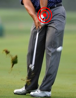
Activity-related pain over the base of the second and third metacarpals should be done in typical evaluation. In golf, baseball, and lacrosse athletes, the pain is typically in the dominant hand and reproduced at the top of the backswing maximal wrist extension; pain can also occur at the point of impact with the ball (e.g., golf, baseball). On physical examination, there may be point tenderness, swelling, and bogginess over the base of the third metacarpal. Pain with resisted wrist extension and passive wrist flexion is suggestive of ECRB insertional tendinitis. MRI scans will show edematous changes to the distal ECRB and its insertion.

The top of the backswing maximal wrist extension in golfer
(Ref: 
https://www.bunkered.co.uk/)


Conservative treatment is the first consideration in the early phase primarily via rest/activity avoidance and use of NSAIDs. Corticosteroid injections can be helpful to reduce inflammation and pain but under caution, as they may lead to tendon attenuation and risk for rupture. Goals for nonoperative treatment need physiotherapy to complete symptom relief and full range of motion by 6 weeks, followed by 2 weeks of gradual strengthening and initiation of sport-specific training around week 12, after the patient’s wrist has reached 85% of the strength of the contralateral side.

Lacrosse
(Ref: https://cmsvathletics.com/)


Patients with mild symptoms or faster progression through this general protocol may return to sports sooner. Tenosynovectomy, the surgery, is indicated after 6 to 12 months of failed nonoperative treatment. Postoperative, patients have their wrists immobilized for 2 weeks, followed by a range of motion therapy. At 6 weeks postoperative, the rehabilitation protocol is the same as the nonoperative treatment described above. 


Fourth-Compartment Syndrome 

The extensor indicis proprius (EIP) muscle originates along the distal third of the ulna and passes within the fourth compartment, deep and ulnar to the extensor digitorum communis (EDC) tendons. 

Extensor Indicis Proprius 
(Ref: 
https://proper-cooking.info/)


Increasing of the space occupied within the fourth compartment was known in term “Anomalous” that can cause pathologic increase in compartment pressure with subsequent tenosynovitis, irritation of the posterior interosseous nerve (PIN), pain, and disability. This is pathomechanics to develop forth – compartment syndrome.

Initial treatment is typically nonoperative, with rest, NSAIDs, activity modification, splinting, and corticosteroid injections. Patients who do not respond to prolonged nonoperative treatment should raise suspicion for the presence of aberrant anatomy (e.g., anomalous muscle or tendon). Surgery is indicated for patients without improvement despite 3 to 6 months of nonoperative treatment, which involves decompression via surgical release of the fourth extensor compartment; concomitant tenosynovectomy and reduction or excision of associated anomalous muscles may be performed to decrease the risk of recurrence, particularly in patients who plan to return to sports. 


Distal posterior interosseous nerve (PIN) Syndrome 

The PIN is the terminal branch of the radial nerve, which passes through the 2 heads of the supinator and travels to the wrist along the radial floor of the fourth extensor compartment, just under the Lister tubercle. Terminal sensory branches of the PIN cross dorsally over the scapholunate ligament and innervate the dorsal capsule of the wrist.

Posterior Interosseous Nerve (PIN)
(Ref: 
https://casereports.bmj.com/content/14/10/e245659)


Athletes whose sports require repetitive, forceful hyperextension of the wrist (e.g., gymnasts, football linemen and defensive backs, platform divers, weight lifters), particularly those with hypermobility at baseline, may experience dorsal wrist pain secondary to impingement of the PIN at the wrist. 

On examination will have pain exacerbated by maximal dorsiflexion of the wrist as well as tenderness localized to the fourth extensor compartment along the course of the PIN. 

Gymnasts on balance bar
(Ref: https://blog.orthoindy.com/)


Initial treatment for athletes with suspected distal PIN impingement is nonoperative by immobilization and NSAIDs. Surgical treatment is indicated when PIN neurectomy has been shown to be a safe and effective procedure for providing pain relief in most patients.


Avascular Necrosis of the Lunate (Kienböck Disease) 

Sometimes, wrist pain is caused by not enough or a blocked blood supply. Presenting symptoms are often similar to those of wrist sprain without a history of trauma. Dorsal wrist tenderness over the lunate with adjacent reactive synovitis and soft tissue swelling is common. Decreased grip strength and pain with motion are usually present and exacerbated by activity, particularly with extension and axial loading across the wrist (e.g., push-ups or military press). 

Military press or overhead press
(Ref: https://www.inspireusafoundation.org/)


Kienböck disease refers to avascular necrosis of the lunate and is the most common type of idiopathic carpal avascular necrosis. Its origin remains unclear and is likely multifactorial, with local vascular and osseous abnormalities being most commonly implicated. It is most common in men aged 20 to 40 years. 

MRI and the presence of uniform signal change of the lunate compared with the rest of the carpus are used for diagnosis. 

Avascular Necrosis of the Lunate (Kienböck Disease) MRI: Black area at carpal
(Ref: 
https://www.orthobullets.com/)


Patient symptoms and radiographic staging of disease are the major treatment guidelines. Symptomatic patients in early stages of disease are typically treated initially with cast immobilization, in order to improve lunate vascularity. In later stages, palliative and performed in an attempt to limit continued carpal collapse (e.g., proximal row carpectomy, wrist arthrodesis, denervation).


Occult Dorsal Carpal Ganglion 

Occult dorsal ganglion cysts may result from athletic activity and lead to a dorsal impingement syndrome. 60%-70% of these mucin-filled cysts originate from the Scapholunate ligament and most commonly present as a cystic mass extruding between the extensor pollicis longus and extensor digitorum communis tendons. 

Smaller, occult dorsal wrist ganglions are more difficult to identify. An inciting injury to the SL ligament and subsequent degenerative change is thought to lead to formation of occult ganglion cysts, although an inciting injury is only reported in about 10% of patients. 

Dorsal Carpal Ganglion
(Ref: https://quizlet.com/)


Diagnosis should be considered for all athletes with dorsal wrist pain that becomes worse with dorsiflexion and loading across the wrist joint. They will have maximal tenderness over the SL interval, which is identified by palpating the soft tissues directly over the inline of the Lister tubercle, which may be exacerbated by passive hyperextension of the wrist.

 Initial treatment is nonoperative, with corticosteroid injection directly into the wrist capsule followed by a period of splint immobilization, which can provide pain relief and help with diagnosis. Surgical intervention is effective for patients with significant activity-limiting pain and nonoperative treatment that has failed. 


Dorsal Capsular Impingement 

Dorsal wrist impingement (DWI) refers to a disorder characterized by mid dorsal wrist pain attributed to capsulitis or synovitis of redundant capsular tissue impinging between the ECRB tendon and dorsal ridge of the scaphoid. The onset may be relatively minor but leads to swelling and thickening of capsular tissue that is prone to recurrent episodes of impingement and a cycle of aggravation with persistent inflammation. 


Dorsal wrist impingement 
(Ref: 
https://journals.sagepub.com/doi/full/10.1177/23259671221088610)


In chronic cases, osteophytes may develop along the dorsal scaphoid, lunate, or dorsal rim of the distal radius, which leads to worsening impingement and dorsal impaction. Pain is localized to the ECRB, where it passes over the dorsal scaphoid, which is exacerbated with full wrist extension and loading of the wrist in an extended position (e.g., tabletop push-off test).

Plain-film radiographs are typically normal, and CT scans may show the development of small osteophytes. MRI scans can be helpful in confirming DWI, which may show dorsal capsular thickening and redundancy with signs of inflammation in this area. 


ECRB anatomy
(Ref: https://www.orthobullets.com/)


The most DWI can be cured by conservative within 2 to 3 months by rest, splint immobilization, and NSAIDs. Corticosteroid injections are helpful to break the cycle of capsular inflammation and swelling and often provide significant (70%) pain relief for several weeks. Surgical treatment may be indicated for refractory cases that fail nonoperative management. 

Operative is needed If conservative treatment cannot solve. Postoperatively, patients are placed in a removable wrist orthosis and begin immediate range of motion therapy, with the goal of full wrist motion at 2 to 3 weeks. Strengthening begins after full motion is achieved, and athletes may begin a return-to-sports protocol around 6 weeks postoperatively, when strength is 80% that of the contralateral side. 


These most anatomy and pathologies have talked about Gymnastic, Lister tubercle, second metacarpal and third metacarpal. The treatment consists of conservative such as immobilize with brace and strengthening therapeutic exercise, and surgery that need post operative rehabilitation.

In fact, there are many wrist disorders syndrome in Sports or daily living that we will discuss together later.


Reference: 

 https://journals.sagepub.com/doi/pdf/10.1177/23259671221088610 

 https://www.ncbi.nlm.nih.gov/pmc/articles/PMC5074830/

 https://www.jospt.org/doi/pdf/10.2519/jospt.2008.2672


วันอาทิตย์ที่ 11 ธันวาคม พ.ศ. 2565

Ligament failure from wrist hyperextension weight bearing basic review


Ref: https://www.naasery.com/

            Have you taken weight on your palm with pain? Taking weight on the palm such as push up, handstand, or hand support is a common function in daily living and sports. To do this function requires wrist extension, that is motion from carpal bones. 

It is FOOSH injury which is nickname for an injury caused by fallen onto an outstretched band.


Different wrist hyperextension weight bearing direction make different ligament injury
(Ref: https://aulakinesica.com.ar/semioquirurgica/files/12389765.pdf)

           Carpal bones are 8 bone components in 2 rows of wrist joint which consists of trapezium, trapezoid, capitate, and hamate in distal carpal, and scaphoid, lunate, and triquetrum in proximal carpal. However, pisiform bone which is sesamoid bone on triquetrum and is easy to palpate because it locates out of carpal line superficially. 

The complex nature of carpal mechanics can be simplified by considering the distal carpal row (trapezium, trapezoid, capitate, and hamate) as securely attached to the medial 4 metacarpals through short, tight, intrinsic ligaments. The distal row moves with the hand as a single unit. The proximal carpal row (scaphoid, lunate, and triquetrum) can be considered a single free-body, intercalated between the hand (including the distal row) and the forearm, suspended by extrinsic radiocarpal and intrinsic intercarpal ligaments. 


Carpal bone anatomy
(Ref: https://www.lecturio.com/)


As the hand forearm unit moves the wrist, the position of the intercalary proximal row shifts at the radiocarpal joint (relative to the forearm) and at the midcarpal joint (relative to the hand), similar to a ball-and-socket joint. The carpal mechanism depends on the health and integrity of the intrinsic and extrinsic ligaments to guide bony relationships among the 7 critical carpals (pisiform excluded). 

Carpal alignment at rest is maintained with considerable stored potential energy and, by definition, a predisposition of the carpus to collapse into a more stable but less physiologic attitude. Ligamentous struts and guy wire mechanisms maintain the longitudinal axis of the scaphoid at about 47 relative to the longitudinal axis of the hand forearm unit. A neutral position of the lunate is maintained through its secure attachment to the proximal scaphoid pole by the scapholunate (SL) interosseous ligament. Separated from the palmar - flexing influence of the scaphoid, the lunate is predisposed to collapse into extension  


Wrist joint component: Radius, Ulnar, Carpals, Metatarsal, and Carpal ligaments
(Ref: https://www.ncbi.nlm.nih.gov/)


    Wrist extension pain is related to carpal instability that can develop to hyperextension. 

Carpal instability can occur after trauma, remotely after trauma, and sometimes without trauma. The 3 most important conditions are scapholunate instability, lunotriquetral instability, and perilunate instability complex. The carpus is considered unstable if it exhibits symptomatic malalignment, is not able to bear loads, and does not have normal kinematics during any portion of its arc of motion.  


There are 4 patterns of instability that have been described by various investigators and are widely accepted Carpal instability dissociative (CID) refers to instability within a row of carpal bones, such as scaphoid fractures or scapholunate dissociation in the proximal row. Carpal instability nondissociative (CIND) is instability between rows either at the radiocarpal joint (eg, radiocarpal fracture dislocations) or midcarpal joint (eg, midcarpal instability). Carpal instability combined/complex (CIC) is a combination of CID and CIND, such as Perilunate dislocations (PLDs). Carpal instability adaptive (CIA) refers to carpal malalignment in adaptation to extra carpal changes (eg, carpal collapse in malunited distal radius fractures). 


Scapholunate instability 


Scapholunate ligament
(Ref: http://rehabforbetterlife.com/)


Scapholunate instability is the most common form of carpal instability. Scapholunate ligament tears wrist instability commonly occurs in a spectrum of severity in hyperextension injuries. Contact sports such as football or rugby commonly place the athlete in a position of impact with hyperextension, ulnar deviation, and supination of the wrist that can lead to these injuries. This may be preceded by a fall. Pain is typically over the dorsum and dorsoradial aspect of the wrist, aggravated by loading the extended wrist or strenuous activity. Pain in a loaded, extended wrist with tenderness in the dorsal wrist at the interval between the third and fourth extensor compartments suggests possible scapholunate interosseous ligament injury. Lab investigations including x - ray and MRI are needed.


Forward wrist weight bearing (hyperextension, ulnar deviation, and supination)
Ref: https://www.amazon.com/)


Stage 1 of 6 which is initial treatment consists of splinting to allow healing in acute and subacute injuries with subsequent proprioception training of the flexor carpi radialis and extensor carpi radialis muscles. Surgery for recalcitrant pain includes arthroscopic interventions, such as de´ bridement alone or with thermal shrinkage and/or pinning, and has 80% to 90% success rates. Left untreated, it may progress to arthritis. 


Lunotriquetral instability 


Lunotriquetral ligament injury develop Lunotriquetral instability
(Ref: https://www.3pointproducts.com/blog/)


Lunotriquetral instability remains frequently underdiagnosed. It may occur in isolation as an acute traumatic tear or in association with degenerative causes, such as ulnocarpal abutment and central TFCC tears, and as part of the perilunate complex injury, to name a few conditions. Acute isolated injuries typically result from a backward fall with the hypothenar eminence striking the ground. Tenderness is localized to the lunotriquetral interval with the appearance of a volar sag, and the Reagan ballottement and Kleinman shear tests may be positive. Stage 1 of 3 which is acute partial injuries, above-elbow casting or splinting with a pad under the pisiform to boost the triquetrum into correct alignment is prescribed. Even in chronic situations, this should be the first line of treatment. Patients who remain symptomatic may benefit from arthroscopic de´ bridement with or without pinning of the lunotriquetral joint. 


Backward fall down
(Ref: https://www.jucm.com/)


Perilunate instability complex 

Perilunate instability complex is the most common wrist dislocation and presents a spectrum of ligamentous and/or bony disruptions to the carpal. The mechanism of injury is wrist axial loading and hyperextension, ulnar deviation, and intercarpal supination. Purely ligamentous injuries are termed lesser arc injuries and those with a fracture greater arc injuries. These terms are synonymous with PLD and perilunate fracture dislocation (PLFD), with the most common fracture a scaphoid fracture. Aside from the acutely painful and swollen wrist, clinical findings may be subtle and there should be suspicion with a high-energy injury mechanism. Acute median neuropathy may be present and was reported in 23% of cases in Herzberg and colleagues’ series. CT scans are indicated when fractures are suspected. This instability may need surgery recommendation from doctors. 


Perilunate instability or dislocation
(Ref: https://link.springer.com/chapter/10.1007/978-1-4471-6572-9_27)


Distal radioulnar joint (DRUJ) instability

    Distal radioulnar joint (DRUJ) instability can occur either from a variety of causes including traumatic and nontraumatic causes. Primary stability of the DRUJ is contributed by the congruity of its articular surfaces and the TFCC. The DRUJ is further reinforced by an osseocartilaginous lip on the volar aspect of the radius. The secondary stabilizers include the joint capsule, extensor carpi ulnaris, pronator quadratus, and interosseous membrane. DRUJ instability can be due to either alterations in the bony anatomy, resulting in altered sigmoid notch architecture or abnormal radioulnar relationship, and/or disruptions to the TFCC. 


TFCC injury develop instability
(Ref: https://pathologies.lexmedicus.com.au/)


DRUJ instability may accompany distal radius fractures due to TFCC disruption or basal ulnar styloid fractures. A fracture of the palmar lunate facet disrupts the buttress effect of the palmar lip and can cause instability. TFCC tears can be traumatic or degenerative. Synovitis in rheumatoid arthritis attrite the stabilizers of the DRUJ, causing instability. 

Patients may present with ulnar-sided wrist pain after a fall on an outstretched hand that is usually exacerbated with loading of the wrist in extension, at the extremes of pronation or supination, or after lifting weights. Patients may report clicking and hypermobility. There may be swelling over the ulnar side of the wrist with a dorsally subluxed ulna head projections of the wrist suspending 2.27 kg of weight and always should be compared with the opposite side. MRIs can delineate foveal and peripheral tears of the TFCC as well as assess the state of cartilage in the DRUJ. 


Basal ulnar styloid fracture is in red box.
(Ref: https://www.reddit.com/r/)


TFCC injuries can occur with injuries to the extensor carpi ulnaris and fractures of the radius and/or ulna bone, and these must be addressed concurrently. Nonoperative treatment of acute TFCC injury involves casting or splinting the patient in the position of stability for a period of 6 weeks. In patients with persistent DRUJ instability after fracture reduction or after a trial of nonsurgical treatment, TFCC repair is warranted. This can be done as an open procedure for arthroscopic assisted or arthroscopic capsular repairs. An open repair can be done via an approach between the fifth and sixth extensor compartments. An inverted L-shaped capsulotomy is made, preserving the dorsal radioulnar ligament. The TFCC is then anchored via bone tunnels or suture anchors.


Triangular Fibrocartilage Complex (TFCC)
(Ref: https://www.orthobullets.com/)


Scaphoid fracture 

Scaphoid fracture type
(Ref: https://www.rch.org.au/)


Scaphoid fractures are the most commonly injured carpal bone with a high incidence in college football players and an increasing incidence in female athletes. This hyperextension wrist injury tends to occur in a pronated, radially deviated hand. Presentation can range from disabling wrist pain to mild swelling and decreased range of motion. It is not uncommon to find a scaphoid nonunion with a remote history of a wrist sprain. Located at the radial side of the carpus, athletes will complain of radial-sided wrist pain with exquisite tenderness in the anatomical snuff box, axial loading of the thumb, or pincer grasp. Radiographic and MRI assessment of the wrist should be involved in diagnosis. 


Anatomical snuff box is represented by red triangle
(Ref: https://teachmeanatomy.info/)


Treatment decisions depend upon fracture location and displacement, with strong surgical consideration being given to scaphoid fractures which are displaced and/or proximal. Whether treatment affects the athlete’s continued participation in his or her sports within the context of the status of the season may also play a role in determining whether or not to operate. Due to retrograde blood supply, distal pole scaphoid fractures can effectively be treated nonsurgical.

There are basic wrist hyperextension injury reviews that raise during wrist extension weight bearing activities. The signs and symptoms are demonstrated on the radial side, the most. Mechanics of injury history, physical assessments, and radiography are combined in diagnosis confirmation. In case of mild damage, it needs to be immobilized by a brace. In case of more instability, it needs to be operative. However, all cases need physiotherapy to improve the quality of movement. 

    
Push up bar
(Ref: https://theworkoutdigest.com/)

                Push up bar or fist weight bearing on thick cushion may compensate pain mechanical.

Ref: https://blog.joinfightcamp.com/


Reference:

http://ortho2.md.chula.ac.th/phocadownload/data-sheet/injuries-wrist-AdisornMD.pdf 


https://josr-online.biomedcentral.com/articles/10.1186/s13018-016-0432-8 


https://aulakinesica.com.ar/semioquirurgica/files/12389765.pdf 


https://www.researchgate.net/publication/319937516_Treatment_of_scapholunate_ligament_injury_Current_concepts 


https://journals.sagepub.com/doi/pdf/10.1177/23259671221088610 


Sports physiotherapy management for tennis elbow and other treatment options.

Ultrasound therapy in tennis elbow treatment (Ref: https://nesintherapy.com/) Tennis elbow is degeneration of the tendons that attach to t...