แสดงบทความที่มีป้ายกำกับ posterior pelvic tilt แสดงบทความทั้งหมด

วันอาทิตย์ที่ 23 ตุลาคม พ.ศ. 2565

Physiotherapy with (second) basic 10 therapeutic exercises in low back pain with flat back patients (ep.2)


Roman chair exercise
(Ref: https://barbend.com/)

      Flat-back syndrome is characterized by forward inclination of the trunk, inability to stand upright, and LBP that decreases the lumbar lordosis of the spine. The decreased lumbar spine lordosis induces changes in absorbing shock between vertebrae, and creating stresses in spinal muscles, tendons and ligaments. The ideal curvature of the spine in the sagittal plane serves to reduce loads on the vertebral discs and any shock to the spine, and it allows effective action of the spinal muscles.


The presence of a flat back is associated with malalignment in the spine, which could cause dysfunction of the deep lumbar muscles and result in chronic low back pain and deep muscle atrophy. Several studies have demonstrated atrophy of the lumbar multifidus muscles with infiltration of fatty tissue in patients with chronic LBP and atrophy at the dysfunctional lumbar level. 


Lumbar erector spinae and multifidus muscles
(Ref: https://centenoschultz.com/)


    The lumbar paraspinal muscles is the neighbor which can be progressively loaded during extension exercises by utilizing back exercise units that will tilt the pelvic to different degrees. As one progresses from a more upright position to a more horizontal position, the exercise becomes more intense for the back extensors. Once a patient can perform the exercise in the prone horizontal position. Sitting extension exercises performed with specialized equipment are also a good way to strengthen the low back musculature, because the resistance can be progressively increased. It has been demonstrated that the exercise activates the low back muscles better if the pelvis is mechanically stabilized so that the extension movement comes from the spine rather than from the hip extensors.


Some studies have shown that in individuals with LBP, moreover, there is also a decrease in the strength and lengthening of the iliopsoas, due to the connection of this muscle with the pelvis and the lumbar spine. As a result of this attachment, the iliopsoas possibly has a stabilizing role in the column. It is thought that tension in this muscle, formed by the union of the psoas major, psoas minor and the iliac acts bilaterally with the insertion, causing an increase in lordosis, whereas weakening of the muscle reduces its size where both these conditions result in pain. 


Force direction of hip flexor (iliacus & psoas) and lumbar back extensor 
(Ref: https://www.sydneyphysioclinic.com.au/)


All of these above muscles are members of pelvic and lumbar stabilizer or core stability muscles. Physiotherapists utilize exercise therapy as an intervention for patients with LBP. The spinal stabilization exercise approach has become popular with many therapists. Physical therapists tend to take different approaches when rehabilitating the muscles of the low back in patients. 


Due to the lumbar multifidus and erector spinae muscles have a relatively high proportion of type I (slow-twitch) muscle fibers. These muscles have fiber composition that makes them well suited for endurance or sustained contraction activities. 


Floor exercise for low back is low impact exercise
(Ref: https://www.popsugar.com/)


There is no evidence in the literature that one exercise program is superior to another. Using low-load stabilization exercise makes them well suited for endurance or sustained contraction activities. So, this topic maintained variation of basic lumbar exercise to treat LBP from flat back posture.  


The second basic 10 of 20 therapeutic strengthening exercises to activate anterior tilt (Remark: If there is tightness of the abdominal muscle or hamstring, it is necessary to treat these muscles to restore normal length before the abdominals can be expected to function optimally.)


Each exercise needs 10 - 15 reps with 3 sets for 3 - 5 days a week.


Exercise #1: Supine hip external rotation with band: exercise with loop band or make band to be loop which need slight heavy resistance. Separate both knees to the floor with slight slow speed both downward and upward directions.


Exercise #2: Heel slide to hip flexion


Exercise #3: Supine hip flexion: In case of too short legs, I would like to recommend you to put some things under your foot at starting position.


Exercise #4: Upper back extension: If you exercise on the bench that will be call Roman chair exercise.

Exercise #5: Seat lumbar extension: If you exercise in fitness, you can exercise with back extension machine.


Exercise #6: Chair pose: During bending the knees, you have to maintain the space between both knees.


Exercise #7: Stand pelvic anterior tilt: Start with bend both knees slightly. Then arch your lumbar spine.


Exercise #8: Lunge: During lunge, do not let knee be inward direction that is keeping knee point to in front. Start with forward - backward stance and move body downward and upward vertically.


Exercise #9: Deadlift: The movement consists of bend knees slightly and straight lumbar with bend over. 


Exercise #10: Unidedal deadlift: It combines of straight lumbar bend over with elevate leg to hip level and opposite hand touch the floor.


The system of local stabilization involves deep intrinsic muscles which are directly attached to the lumbar vertebrae, and the global system comprises the great superficial muscles originating in the pelvis which insert in the thoracic cavity, with both systems necessary for stability and control of movements.


The erector spinae and multifidus muscles are the primary muscle groups responsible for controlling lumbar motion and forward inclination of the trunk. It is estimated that the erector spinae and multifidus contribute up to 85–95% of extensor moment during manual handling tasks, with these muscles playing an important role in resisting anterior shear forces during lifting and lowering.
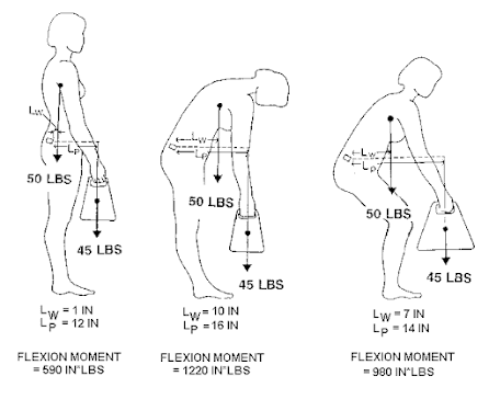

Load on lumbar spine in different lifting posture
(Ref: https://ouhsc.edu/)


The erector spinae and multifidus muscles are thought to play an important role in the prevention of back injuries, and these muscles are often targeted during the rehabilitation of patients with such injuries. For example, during vocational activities such as lifting, the erector spinae and multifidus muscles are the major contributors to the extensor moment and serve to resist anterior shear forces acting on the lumbar spine.


Core stabilization exercises aim to maintain this stability, improve strength, resistance, improving neuromuscular control  of the abdominal and lumbar muscles, and attenuating recurrent LBP. Stabilization exercises are essential in order to provide a base for movement of the arms and legs when supporting weight and to protect the medulla and spinal nerves. Exercise stabilization programs emphasis on the transverse abdominis and multifidus (deep trunk muscles). Paravertebral and abdominal muscles such as the pelvic musculature and the diaphragm are also important targets for exercise. 


Upright with torso bending contributes increased lumbar disc pressure and multifidus workload
(Ref: https://ergonomictrends.com/)


80% to 90% of patients with acute LBP seem to recover within 6 weeks, regardless of the treatment received. In spite of this, there is about a 60% recurrence rate of LBP in patients within 1 year of the initial episode. Some patients do not recover from the acute LBP episode and go on to have a chronic condition. LBP is one of the leading causes of incapacity and the high cost of treatment renders preventative strategies paramount. Therefore, proper LBP prevention and treatment can help you to maintain daily life living capacity and save money. 


Lifting stuff properly is one of low back pain prevention
(Ref: https://reliva.in/)


Reference

Kendall FP., et al. Muscles testing and function. Fourth edition. Williams & Wiikins. USA.

1993.  


https://www.researchgate.net/publication/258825540_Effect_of_Individual_Strengthening_Exercises_for_Anterior_Pelvic_Tilt_Muscles_on_Back_Pain_Pelvic_Angle_and_Lumbar_ROMs_of_a_LBP_Patient_with_Flat_Back


https://www.mdpi.com/1660-4601/18/20/10923/htm 


https://www.ncbi.nlm.nih.gov/pmc/articles/PMC5342962/ 


https://www.scielo.br/j/rbfis/a/HjjyDzqVhbvDxkCSHqnWkjs/?format=pdf&lang=en


 https://drrobertlaprademd.com/wp-content/uploads/2015/07/rehabilitation-exercise-progression-for-the-gluteus-medius-muscle-2011.pdf 


https://www.researchgate.net/publication/333795080_Evaluation_and_comparison_of_electromyographic_activity_in_bench_press_with_feet_on_the_ground_and_active_hip_flexion 


https://www.scielo.br/j/fm/a/z6pw7PhWLtMQDGWyZCmYP7c/?lang=en&format=pdf 


https://www.researchgate.net/publication/272516106_The_Effectiveness_of_Lumbar_Extensor_Training_Local_Stabilization_or_Dynamic_Strengthening_Exercises_A_Review_of_Literature


https://paulogentil.com/pdf/The%20progression%20of%20paraspinal%20muscle%20recruitment%20intensity%20in%20localized%20and%20global%20strength%20training%20exercises%20is%20not%20based%20on%20instability%20alone.pdf 


https://www.jospt.org/doi/pdf/10.2519/jospt.2008.2865


https://www.researchgate.net/publication/285939823_Influence_of_Stretching_and_Strengthening_of_the_Iliopsoas_Associated_with_Lumbar_Segmental_Stabilization_Exercises_in_Patients_with_Low_Back_Pain_The_pilot_study 


วันเสาร์ที่ 1 ตุลาคม พ.ศ. 2565

Physiotherapy with basic 9 stretching to improve low back pain in flat back syndrome

Flat back posture
(Ref: https://www.orthobullets.com/)


            I have taken care of some patients who had low back pain which is associated with decreased lumbar spine lordosis. Some of them got pain from sports activities such as cycling in triathlons.

    The decreased lumbar spine lordosis or hypo-lordotic lumbar curve consists of posterior pelvic tilt that ASIS level is same or higher than PSIS level, and lumbar spine trend to straight that means flexion. 


    We have seen the hypo-lordotic lumbar curve in common two posture including 

(1) Sway - back posture, the lower component illustrates lumbar spine flexion (flattening) of lower lumbar area, pelvis posterior tilt, hip joints hyperextended with anterior displacement of pelvis. Because we focus on tightness of structure in this article, the muscle shortness consists of hamstrings, upper fibers of internal oblique, and low back muscles.


Dark gray area represents muscle weakness in sway - back posture
(Ref: https://www.pinterest.com/)


(2) Flat - back posture, the characteristic of this posture associated with lumbar spine flexed (straight), pelvis posterior tilt, hip joints extension. The muscle shortness has been found at the hamstring, but abdominal muscles are strong that may or may not shortness. Sometimes, Gluteus maximus is included as well. 


Dark gray area represents muscle weakness in flat - back posture
(Ref: https://www.pinterest.com/)


Both of them have hamstring shortening where hamstring anatomy attachment is at the ischial tuberosity, and this structure moves superiorly as the pelvis tilts. Hamstring tightness is a common finding in the LBP patient. 

I have found not only all muscles as above, but also both quadratus lumborum (QL) in low back pain with lumbar hypo-lordotic. They are taut and trigger both sides so that they are not improved by massage and stretching. I have skipped massage and stretching on QL and applied another technique to release them. Moreover, I have always seen TFL tightness in flat back posture more than hyper lordosis posture. 


Muscles around pelvic that hamstring and gluteus maximus are the key of tightness
(Ref: https://musculoskeletalkey.com/)


The principle to stretch this muscle is the same as the others: stretch to the point where “tightness with pain” or “noticeable tension without pain” will hold at the point for 30 seconds of 3 - 5 reputations following demonstrated VIDEO. 


Basic 9 stretching to correct posterior pelvic tilt. (Please stop stretching and consult a physiotherapist or doctor if you feel worse pain and/or numbness.)

Exercise #1: Stand toe touch double legs: Keep back straight and/or pelvic anterior

tilt together with knee straight during stretch. Reach as far as you feel some tension

that means no need to push excess muscle length. Your range of motion will increase

gradually in the future.


Exercise #2: 90-90 SLR with knee straight: Straight target knee as far as you feel some

tension that means no need to push excess muscle length. Your range of motion will

increase gradually in the future.


Exercise #3:  90-90 SLR with knee straight with belt assisted: Put the belt at the ball of foot.

Straight target knee as far as you feel some tension that means no need to push excess

muscle length. Your range of motion will increase gradually in the future. 


Exercise #4: Doorway with knee straight: Keep back straight and/or pelvic anterior tilt 

together with knee straight during stretch. Reach as far as you feel some tension that

means no need to push excess muscle length. Your range of motion will increase

gradually in the future. You can apply it with a table, chair, fence, or any stable

structure as well.


Exercise #5:Supine cross leg: Do not lift shoulder and pelvic from the floor during stretching.

For more stretch, pull the knee to the opposite shoulder.


Exercise #6: Supine figure 4: Keep back straight during stretching to prevent back pain.

For more stretch, keep the knee and chest closer. 


Exercise #7: Basic Cobra with lateral bending stretch: During stretching needs to keep

pelvic on the floor.


Exercise #8: Modified Cobra with lateral bending stretch: During stretching needs to keep

pelvic on the floor.


Exercise #9: Seat torso rotation stretch.


 The flat back,  as the name implies, is a type of sagittal imbalance in the spine, and it is characterized by loss of the lordotic curve. There is a straight back in both the lumbar and thoracic areas except that there is some degree of flexion in the upper thoracic area that accompanies the forward head position. 

The flexible flat - back posture appears to be more common among certain cultures than among others. Asians seem to exhibit this type of posture more frequently than most Americans or Europeans. This type of flat - back does not give rise to as many problems of low back pain as does the lordotic back, or the sway - back posture. The range of motion in extension is usually normal and may be excessive.


Passive lumbar extension test
(Ref: https://www.uma.es/)


Although the pathogenic mechanism remains unclear, poor posture and lack of exercise are major causes of spinal deformity. The normal curve of the spine is known to have a buffering effect against gravity and provides optimal, coordinated body movements. An abnormal spinal curvature is associated with musculoskeletal and nervous systems problems.

The flexible functional state impairment of muscles in the flat-back posture demonstrates gluteus maximus is shortened and hypoactive; in hamstring parts are also shortened yet hyperactive.

90-90 SLR test for hamstring tightness
(Ref: https://bendandmend.com.au/)

The sway - back posture is one in which there is a posterior displacement (swaying back) of the pelvis. The pelvis is in posterior tilt and sways forward in relation to the stationary feet causing the hip joint to extend. The effect is equivalent to extending the leg backward with the pelvis stationary. With posterior pelvic tilt, the lumbar spine flattens and hence there is no lordosis although the long curve in the thoracolumbar region (which is due to the backward deviation of the upper trunk) is sometimes mistakenly referred to as a lordosis. (the term sway - back posture is an appropriate label and requires that the word “sway - back” not be used synonymously with the word “lordosis”)

Pelvic in sway back posture is in front of torso
(Ref: https://ebrary.net/)


In the past, the words “lordosis” and “sway - back” were used interchangeably in referring to the curvature in the low back and lower thoracic arrears. The postural differences between the lordosis and the sway-back postures were recognized in posture and pain. Separating the use of these terms also differentiated these two postures. They are distinctly different with respect to the anteroposterior tilting of the pelvis, the position of the hip joint, and the accompanying muscle imbalance that exist.


Functional state of muscles flexibility deficit in the sway-back posture show the shortened

muscles are suboccipital, sternocleidomastoid, scaleni, chest muscles—pectoralis major and minor, erector spinae lumbar part (lower part), upper fibers of abdominal muscles, gluteus maximus, and hamstrings. All these muscles demonstrate hyperactivity (except for the lower part of lumbar erector spinae, posterior part of internal oblique abdominal muscle, gluteus maximus, and hamstrings.


Gluteus maximus muscle length assessment 
(Ref: https://www.physio.co.uk/)


Clinically, often assume that tighter hamstring muscles will limit anterior tilt of the pelvis. It has been argued that anterior tilt of the pelvis will be reduced because the hamstrings attach to the ischial tuberosity, and this structure moves superiorly as the pelvis tilts, increasing the distance between the attachments of the hamstrings. However, the line of action of the hamstrings is almost vertical, and the attachment to the ischial tuberosity is only slightly posterior to the femoral head. 

This minimal posterior force tending to posteriorly rotate the pelvis is likely to be outweighed by activity of the hip flexors tending to anteriorly rotate the pelvis. Therefore, any change in the length of the hamstrings may not alter the total range of pelvic tilt. The study has shown that no association exists between hamstring muscle length and total range of pelvic motion during forward bending in asymptomatic subjects.


Pelvic posterior tilt force direction
(Ref:https://healthfitinc.com/)


Although hamstring stretching may not affect pelvic tool posture, stretching the short hamstring muscles to correct the faulty alignment is recommended strongly. Short hamstring muscles, because of their attachments to the posterior leg and to the ischial tuberosity, may limit hip flexion ROM. The restricted hip motion is coupled with excessive lumbar motion. Stretching short hamstring muscles to increase hip flexion, therefore, may affect lumbar motion during forward bending. Excessive lumbar motion would increase tensile loads on the spine and thus could result in LBP. The short hamstring muscles cause back pain due to their influence on lumbopelvic rhythm during forward bending. 


SRL stretch
(Ref: https://www.hss.edu/)


Based on the studies described, standing lumbopelvic posture has not been clearly related to muscle length. Furthermore, there is insufficient data to determine the effect of hamstring muscle stretching on standing lumbar and pelvic posture or the relative amounts of lumbar and hip motion during forward bending. Stretching was performed with the patients positioned supine by actively extending the knee with the hip flexed. The SLR measurement method was used first, followed by the AKE method. The results of this study suggest that there is no relationship between hamstring muscle length and lumbopelvic posture. There was some indication, however, that stretching the hamstring muscles may affect motion during forward bending. The data gathered in this study suggest that a 3-week program of hamstring muscle stretching (1) will not alter standing lumbar and pelvic postures, (2) will produce greater forward bending as a result of increased motion at the hips, and (3) may alter the pattern of lumbar and hip motion during forward bending. 


Stand forward bending stretch
(Ref: https://www.verywellfit.com/)


One case study report suggested the lumbar extension traction which is used to restore lordosis to the elliptical normal L1–L5 of the absolute rotation angle value of −40°. It was used for treatment as a part of the chronic low back pain rehabilitation program resulting in large increases in sagittal lumbar lordosis, decreased anterior sagittal balance and a minimization of pain levels in two patients diagnosed with FBS suffering from chronic low back pain. Two chronic low back pain with flat back posture patients in this study demonstrated improvement significantly after intervention at least 16.5 weeks.

Biomechanically, lumbar extension traction increases lordosis due to lumbar extension traction creating a deformation in the soft tissues (muscles, ligaments, and discs) of the lumbar spine. Tendons, ligaments, and discs, all display visco-elastic properties. When the soft tissues of the spine are subjected to a sustained load for a given time, these tissues undergo two major processes, creep and stress relaxation. Creep is the amount of deformation occurring in the tissues and stress relaxation is a reduction in the amount of the internal stress found in the tissue over time. 


Creep phenomenon graph
(Ref: https://web.iit.edu/sites/web/files/departments/academic-affairs/academic-resource-center/pdfs/MaterialsCreep.pdf)


In extension creep loading of cadaveric lumbar specimens, during the first 5 minutes much of the initial deformation is recoverable strain energy (elastic). Most of the non-recoverable strain (permanent deformation) energy takes place from 5 to 20 minutes; at 20 minutes a plateau effect takes place. It is the non-recoverable strain energy that results in the permanent deformation or resting length change of the spinal tissues. The two patients in this study performed extension traction for 20 minutes to take advantage of the visco-elastic deformation in the spinal tissues.


If this type of posture exists without low back pain, it is not necessary to change it. If the back is painful, and restoring the normal anterior curve is indicated.


Reference :

http://www.norrishealth.co.uk/library/sdarticle.pdf


https://d-nb.info/1166557340/34


https://www.mdpi.com/1660-4601/18/20/10923/htm 


https://watermark.silverchair.com/ptj0836.pdf?token=AQECAHi208BE49Ooan9kkhW_Ercy7Dm3ZL_9Cf3qfKAc485ysgAAAsMwggK_BgkqhkiG9w0BBwagggKwMIICrAIBADCCAqUGCSqGSIb3DQEHATAeBglghkgBZQMEAS4wEQQMjEMqUKs03vtPoJ-fAgEQgIICdtNoCBekqxzlyP7XaRxkJDjk82KXkEGVKY2R090TUzcP0c6NFC1oowVjkl_LvNhGk1daUTmmCImrhrhVDPbNhMTl4gz-1H87fF3Zw42eNjQvuv3ELCbOISwPxPTKzrusg0kk-R4FrHtudhiw-dv7d00eS46T73OFjvPhd1MbiSAow_kqRXUzcFMTWsPfn0heEFEhyWG20mfa-PKaeEWtLgJIVV7iWAu-0Tv4x2JxKp7hGF4zUGZls4QrDQvWz95VkrsrbW8dvOO0iN2ocr_PrInxrgFQXp7thYClvmXySqOH7T-5Lg8aC43X2gJjmufTDHE-Yu9l1kZhcpvdUnNih509fufqNwZk21gLkD5aFNNNcvkF9Qsw0YhDc74zlYQie4oLZvPoi2RvFBIl1mTo3GjAD6POQeaG_uGfViMaNdC4BlTNrSK9dZhX2FngHt7SvwQ8WJQUNDhHk0h78qjQh4t0TDA6uwHGBL7vXU8h6A1169_BC4Qbugw7bKiXEoC37PMhe7cSJgFl61yYaKiKmscKnxIiOnSG0ALEwkU0Mgu2Xpy1jXBNiOnJkUb3Ch-npQV1Q6XKhUjbVOgsGWz4sHYHC0vSNHxwWf9fuELpOqHidAnjh5em27yt7CNB-KFvlAPrG6PV8fXLN0Ild8ATDYwAtbW_VS0VT4nelUDl5vmY-qDC5Xxg1WTr7XA0xk5yYbuBJnJ2x0PLuqrQwkP-ZM8qbRmynczNFpMazJasQ_pdl_1d1Mkcnc3caVLqyWtImXFU8Zull9-CChnOlIVk_QHd4rkWmp7anK3lIypT_Z-z6coSFeccRJg7zf0CpXPGbj3k23OMmQ 


https://www.researchgate.net/publication/326852561_Non-operative_correction_of_flat_back_syndrome_using_lumbar_extension_traction_a_CBPR_case_series_of_two


https://www.ncbi.nlm.nih.gov/pmc/articles/PMC6110233/


Kendall FP., et al. Muscles testing and function. Fourth edition. Williams & Wiikins. USA.

1993.  


Sports physiotherapy management for tennis elbow and other treatment options.

Ultrasound therapy in tennis elbow treatment (Ref: https://nesintherapy.com/) Tennis elbow is degeneration of the tendons that attach to t...