 |
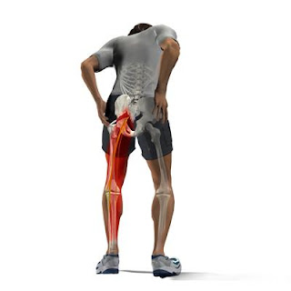
| Hamstring strain in rugby players (Ref: https://nicksportphysio.wordpress.com/hamstring-strain/) |
The evidence on how hamstring complex derived this name originates from the early Germanic language as well as the butchery trade. Slaughtered pigs were hung from these strong tendons, hence the reference to ‘ham’ (meaning ‘crooked’ and thus referring to the knee, the crooked part of the leg) and ‘string’ (referring to the string-like appearance of the tendons).
Hamstring injuries are one of the most common problems in sports medicine due to athletes’ absence in games. Their prevalence is estimated to reach 12–15% among professional football players. It is also a major problem of track and field sports, dancing, and skiing.
 |
| Ref: https://bodyfocusphysioclinic.com.au/risk-factors-for-hamstring-strain/ |
As well as, hamstring tightness was suspected to be the cause of low back pain, although, not absolutely clear. It has been found hamstring tightness in low back pain patients. Asymptomatic low back pain was better for hamstring flexibility than symptomatic significantly. However, Hamstring tightness had no influence on pelvic motion in both groups during forward bending.
 |
| Sacrotuberous ligament play a role as the junction of hamstring and lower back (Ref: https://www.clpt.fit/blog/) |
The important component of hamstring stretching needs a full straight leg and simultaneously bending forward at the groin without spine bending. To stretch in a position of anterior pelvic tilt results in a significantly greater increase in hamstring flexibility.
 |
| Left: correct hamstring stretching with spine straigth Right: incorrect hamstring stretching with spine flexion (Ref: https://www.sulmall.com/?category_id=6116823) |
15 ways to stretch hamstring
Exercise #1: Hamstring half wall stretch:Keep back straight and/or pelvic anterior tilt during stretch. You can apply with wall, chair, or any stable structure as well.
Exercise #2: Sit and reach double legs: Keep back straight and/or pelvic anterior tilt together with knee straight during stretch. Reach as far as you feel some tension that means no need to push excess muscle length.
Exercise #3: Sit and reach single leg: Keep back straight and/or pelvic anterior tilt together with knee straight during stretch. Reach as far as you feel some tension that means no need to push excess muscle length. You range of motion will increase gradually in the future.
Exercise #4: Stand toe touch double legs: Keep back straight and/or pelvic anterior tilt together with knee straight during stretch. Reach as far as you feel some tension that means no need to push excess muscle length. You range of motion will increase gradually in the future.
Exercise #5: Stand toe touch single leg: Keep back straight and/or pelvic anterior tilt together with knee straight during stretch. Reach as far as you feel some tension that means no need to push excess muscle length. You range of motion will increase gradually in the future.
Exercise #6: Stand hamstring stretch: Keep back straight and/or pelvic anterior tilt together with knee straight during stretch. Reach as far as you feel some tension that means no need to push excess muscle length. You range of motion will increase gradually in the future. You can apply with table, chair, fence, or any stable structure as well.
Exercise #7: 90-90 SLR with knee bend: Straight target knee as far as you feel some tension that means no need to push excess muscle length. You range of motion will increase gradually in the future.
Exercise #8: 90-90 SLR with knee straight: Straight target knee as far as you feel some tension that means no need to push excess muscle length. You range of motion will increase gradually in the future.
Exercise #9: 90-90 SLR with knee bend with belt assisted: Put the belt at ball of foot. Straight target knee as far as you feel some tension that means no need to push excess muscle length. You range of motion will increase gradually in the future.
Exercise #10: 90-90 SLR with knee straight with belt assisted: Put the belt at ball of foot. Straight target knee as far as you feel some tension that means no need to push excess muscle length. You range of motion will increase gradually in the future.
Exercise #11: Seat toe touch: Keep back straight and/or pelvic anterior tilt together with knee straight during stretch. Reach as far as you feel some tension that means no need to push excess muscle length. You range of motion will increase gradually in the future.
Exercise #12: Seat and reach: Keep back straight and/or pelvic anterior tilt together with knee straight during stretch. Reach as far as you feel some tension that means no need to push excess muscle length. You range of motion will increase gradually in the future.
Exercise #13: Step toe touch: Keep back straight and/or pelvic anterior tilt together with knee straight during stretch. Reach as far as you feel some tension that means no need to push excess muscle length. You range of motion will increase gradually in the future.
Exercise #14: Doorway with knee bend: Keep back straight and/or pelvic anterior tilt together with knee straight during stretch. Reach as far as you feel some tension that means no need to push excess muscle length. You range of motion will increase gradually in the future. You can apply with table, chair, fence, or any stable structure as well.
Exercise #15: Doorway with knee straight: Keep back straight and/or pelvic anterior tilt together with knee straight during stretch. Reach as far as you feel some tension that means no need to push excess muscle length. You range of motion will increase gradually in the future. You can apply with table, chair, fence, or any stable structure as well.
Hamstring is one of postural muscles that its tightness may be linked to postural disturbances by anatomy. The sacral nodular ligament is located between the sacrum and the ischial tuberosity, and is present in one fascial line along the erector spinae and occipital bone to the orbital ridge, and to the all posterior section of leg, including hamstring. Decreased hamstring flexibility causes non-specific low back pain or changes in lumbar pelvic rhythm that develop the posterior inclination or pelvic rotate backward. Pelvic rotation backward is one component of abnormal posture that can be found in swayback, flatback syndrome, kyphosis, lordosis, and scoliosis. This mechanics provides tension in the spinal column, sacrum fascia, ligaments and joint pockets, and a limitation of pelvic mobility due to tension in the hip flexors and extensors, which can be considered as a major cause of low back pain. Through this, self-stretching of the hamstrings can be expected to have a great effect when the pelvis is inclined posteriorly.
However, stretching in patients with lumbar disc herniation and sciatic irritation should be very careful and under supervision from doctor and physiotherapist.
The hamstring complex is a biarticular muscle group which works by flexing the knee and extending the hip. Many movements in daily living function need hip flexion and knee flexion at the same time, with opposing effects on hamstring length. The most common modifiable factors are imbalance of muscular strength with a low hamstring to quadriceps ratio (H:Q ratio), muscle fatigue, hamstring tightness, insufficient warm up, and previous injury.
 |
| Lower right: terminal swing phase of running that is initiation of foot contact (Ref: https://doctorlib.info/anatomy/running-anatomy/3.html) |
The most common two specific mechanisms are described for hamstring injuries from daily living function. First, during the terminal swing phase of running, the hamstrings absorb elastic energy to contract eccentrically and promote deceleration of the limb’s advance in preparation for the initial contact of the calcaneus. In this phase, muscles become more susceptible to damage;the biceps femoris muscle is the most affected, as it is more active than the semitendinosus and semimembranosus muscles.
Second, the common damage to the proximal portion of the semitendinosus muscle is a movement of combined high power and extreme range of hip flexion with knee extension, which biomechanically matches the movements of kicking, running hurdles, and artistic dancing.
 |
| Ref: https://www.offset.com/photos/mixed-race-woman-kicking-soccer-ball-68325 |
Most rehabilitation programmes are based on the tissue’s theoretical healing response that include 5 phases. In the acute phase which is the first one needs only rest, ice, compression, and elevation to control hemorrhaging and minimize inflammation and pain. Step to the third phase which may be 1 - 6 weeks after injury the inflammation signs are resolved, hamstring muscle becoming less flexible. This is probably due to pain, inflammation, and connective tissue scar formation, therefore, hamstring stretching can begin.
Hamstring located at the back of the thigh that consists of 4 members of the hamstring family. The three proximal attachments of the hamstrings that include the semitendinosus (ST), long head of the biceps femoris (long head, lhBF) and semimembranosus (SM) muscles originate from the ischial tuberosity.
 |
| Inner thigh to outer thigh muscles: Semimembranosus - Semitendinosus - Bieceps femoris (Ref: https://www.istockphoto.com/) |
(1) The semitendinosus muscle (ST)
It lies in the posteromedial area of the thigh. The ST and lhBF have a common origin on the posteromedial aspect of the ischial tuberosity. The fusiform shape (from external aspect) and has a characteristic oblique or V-shaped raphe (tendinous inscription), runs distally and medially from its proximal insertion on the ischial tuberosity and lies directly on the SM. From its origin, the ST creates a conjoined tendon with the lhBF forming an aponeurosis. The distal tendon starts below the mid-thigh and runs around the medial condyle of the tibia to its distal insertion as a part of pes anserinus. It also unites with the tendon of gracilis and gives off a prolongation to the deep fascia of the leg and the medial head of gastrocnemius.
 |
| Ref: https://learnmuscles.com/glossary/semitendinosus/ |
(2) The semimembranosus muscle (SM)
This muscle is the largest of all of the hamstrings. The SM origin is separate from the ST and lhBF that is located anterolaterally from the ST/lhBF attachment. Fibers of the proximal SM attachment are twisted before forming a proper tendon. It lies posteromedially in the thigh and has a similar location as the ST. It starts on the anterolateral part of the ischial tuberosity to the medial condyle of the proximal tibia to the pes anserinus and descends under the ST, from its wide proximal insertion. The proximal and distal tendons of SM overlap. It means that the part of fibers in the middle of SM has a connection to both tendons: the proximal and the distal. Its anatomical variation attachment may extend to the coccyx or have slips that join with the tendon of adductor magnus.
 |
| Ref: https://www.sciencephoto.com/ |
The biceps femoris muscle (BF) forms the posterolateral part of the thigh. It consists of two heads:
(3) The long head of the biceps femoris (lhBF)
This muscle is of particular interest given its susceptibility to injury. Its unique muscle architecture and the arrangement of its proximal tendon which it shares with semitendinosus, a feature which may explain why injuries to these two muscles can occur simultaneously. It has a common origin on the posteromedial aspect of the ischial tuberosity. The proximal tendon runs laterally after division of the conjoined tendon with the ST. Like in the SM, the proximal and distal tendons of the lhBF are overlapping.
The insertion tendon attachment divides around the lateral collateral ligament, forming two tendinous and three fascial components. Tendinous insertion is into the lateral and anterior aspects of the fibular head and the tibial plateau, while the fascial components mainly attach both heads to the lateral collateral ligament
 |
| Left: long head of biceps femoris muscle Right: short of biceps femoris muscle (Ref: https://learnmuscles.com/glossary/biceps-femoris/) |
(4) The short head of the biceps femoris (shBF).
The proximal attachment of short head of biceps femoris (shBF) arises on the middle third of femur. Its origin is located on the lateral lip of the linea aspera, descending distally and laterally.The shBF originates in the posterolateral region of the femur. It fuses with the lhBF in the distal part of the thigh, forming an aponeurotic structure. The conjoined distal tendon of both heads attaches to the head of the fibula.
The ischial tuberosity is also the area of the distal attachment of a sacrotuberous ligament (STL). This ligament is an elastic and dynamic structure that fibers are descending from the sacrum to the ischial tuberosity in continuity with fibers of the lhBF.
 |
| Red circle illustrated sacrotuberous ligament (Ref: https://www.rehabcareclinic.com/) |
The main function of hamstring muscle is knee flexion and hip extension. For individual function outcomes the contraction of the BF rotates the tibia and fibula externally. Consequently, it prevents internal rotation of the tibia in relation to the femur. The BF is the most effective hamstring muscle in reducing the ACL-loading component produced by the QF through decreasing anterior tibial translation. The ST and SM contraction induce an internal rotation of the tibia. These muscles are antagonists of the external rotation generated by the BF.
The principle to stretch this muscle is the same as the others: stretch to the point where “tightness with pain” or “noticeable tension without pain” will hold at the point for 30 seconds of 3 - 5 reputations following demonstrated VIDEO. However, strengthening the hamstring both of concentric and eccentric contraction are still necessary in prevention and treatment.
Reference:
http://robertsmigielski.pl/wp-content/uploads/2020/04/art1.pdf
https://www.aspetar.com/journal/upload/PDF/2013121863735.pdf
https://www.ncbi.nlm.nih.gov/pmc/articles/PMC1725237/pdf/v039p00319.pdf
https://www.scielo.br/j/rbort/a/XFWsjTdV97KvRXdCX3CCkzQ/?lang=en
Park D. S., Jung S. H. Effects of hamstring self - stretches on pelvic mobility on persons with low back pain. Phys Ther Rehabil Sci 2020;9(3):140 - 148.


