 |
| Ref: https://dailycannon.com/ |
Calf muscles are on hind - lower leg, it plays an important role in daily living and sports activities. Their main function is plantar flexion at the ankle joint where gastrocnemius and soleus are prime mover, and are associated by tibialis posterior, Flexor Hallucis Longus, Flexor Digitorum Longus, peroneus longus and brevis. They also move their ankle and foot more than one direction in one time and support their feet during walking or running or jumping.
In my sports physiotherapy experience, my patients with shin splints, achilles tendinitis, and muscle cramp were treated by stretching. Stretching is one part of physiotherapy treatment that needs appropriate timing and intensity. Stretching is not proven to prevent sports injury because they need several factors e.g. strength, footwear, floor surface, sports specific skill. At least, stretching for flexibility can help to decrease the chance of muscle cramp.
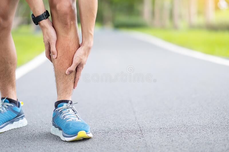 |
| Shin Splints pain (Ref: https://www.dreamstime.com/) |
Knee and toe position in stretching are determined by anatomy. To stretch gastrocnemius need to keep the knee straight because it covers the knee and ankle joint. For soleus which is one joint muscle that covers only the ankle joint, so it needs to stretch with knee bending for maximum result. FHL and FDL run between below knee joint to toes that can be stretched by knee and toe bending.
9 Options to stretch calf muscle
Exercise #1: Sit gastrocnemius stretch with belt assist: Put belt at ball of foot and pull the belt during knee straight.
Exercise #2: Sit soleus stretch with belt assist: Put belt at ball of foot and pull the belt during knee bending.
Exercise #3: Wall stand gastrocnemius stretch: Put hands on the wall for balance with step target leg backward in knee straight position. Then, move pelvic forward with keep heel on the floor.
Exercise #4: Wall stand soleus stretch: Put hands on the wall for balance with target foot is somewhat distance away from the wall. Bend target knee to the wall with keep heel on the floor.
Exercise #5: Soleus chair stretch: Place target foot on the firm cushion chair. Lean body weight forward with keep heel on the chair.
Exercise #6: FHL and FDL chair stretch: Place target foot on the firm cushion chair with toes on roll towel. Lean body weight forward with keep heel on the chair.
Exercise #7: Peroneus ankle inversion stretch: Roll target foot outward as ankle inversion on the floor during sit on the chair. Put other foot a bit above target ankle joint and push.
Exercise #8: Heel drop gastrocnemius stretch: This stretching needs stable step or stairs. Place ball of foot on the edge of stairs and drop target heel down with keep knee straight during stretch.
Exercise #9: Calf stretch board: in my experience, it is the best for stretch with knee straight. You needs hands holding with stable structure for balance.
The members of calf or posterior compartment muscle family
1. Gastrocnemius
Gastrocnemius is one of the triceps surae groups and the other is soleus. The gastrocnemius is more superficial than soleus and covers it. It is a fusiform muscle which consists of two heads which originate from the posterior medial and posterior lateral metaphyseal areas of the distal femur. The distal attachment is variable as the fibers intermingle for a two to three centimeter length before becoming indistinguishable. It maintains a separate entity from the soleus for the length of the muscle belly and only connects into the soleus aponeurosis distally after the gastrocnemius aponeurosis becomes tendinous only.
It spans two joints and is therefore not only a plantiflexor at the ankle but also an assistant flexor of the knee. The effect of the gastrocnemius on the tension of the achilles insertion is dependent on the knee position. Knee extension is required for maximal tension.
 |
| Gastrocnemius muscle (green) covers soleus muscle (red) (Ref: https://www.kenhub.com/en/library/anatomy/gastrocnemius-muscle) |
2. Soleus
Soleus is one of the triceps surae groups and the other is gastrocnemius. The soleus lies deep to the gastrocnemius. It is of pennate shape and a large flat muscle that was given its name from its resemblance to the sole. The origin of this muscle is the oblique line and the middle third of the medial border of the tibia, from a fibrous arch between the fibula and the tibia and from the posterior surface of the head of the fibula. The soleus and gastrocnemius muscles meet in the achilles tendon so that the anterior fibers of the soleus run further downwards along the tendon and the posterior proximal metaphyseal diaphyseal area of the tibia. The strong Achilles tendon inserts at the posterior surface of the calcaneus.
It acts to plantarflex the ankle joint via its conjoint tendon because it locates below the knee joint.
 |
| Soleus muscle (right) (Ref: https://www.shutterstock.com/search/soleus) |
3. Achilles tendon
The Achilles tendon is the thickest and strongest tendon in the human body. Referring to the term of Achilles, Achilles who was the ancient Greek hero of the Trojan war that gave his name to the Achilles tendon. In the story, Achilles was the son of Thetis the nymph who tried to make him immortal by dipping him in the river Styx. However, he was left vulnerable at the part of the body she held him by at his heel. He was killed by an arrow to his heel which was a particularly vulnerable point.
The achilles tendon is surrounded throughout its length by thin gliding membranes called the paratenon. The paratenon functions as an elastic sleeve (although probably not so effectively as a true tendon sheath) and permits free movement of the tendon within the surrounding tissues. A bursa lies between the upper part of the calcaneus and the anterior surface of the achilles tendon.
It is located in the posterior superficial compartment of the lower leg. The origin begins near the middle of the calf fusing with the gastrocnemius muscle proximally. It is broad close to its origin and receives muscle fibers from the soleus almost to its lower end. So, the origin of the achilles tendon is the insertion of the gastrocnemius and the soleus.
The configuration of achilles tendon insertion is broad at its origin, gradually becoming thinner at the midsection. Then, it becomes more rounded until approximately 4 cm above the calcaneus before expanding. The end attachment insertion to the midpoint of the posterior surface of the calcaneus.
Because the insertion at the calcaneus is medial to the axis of rotation of the subtalar joint, the triceps surae is also a weak invertor of the foot.
 |
| Achilles tendon (White) (Ref: https://www.medi.de/) |
4. Tibialis Posterior
Tibialis posterior is a member of the calf muscle group which is located in deep muscle in the calf region and plays many roles including ankle inverter, adductor, supinator, plantar flexor, and foot supporter in walking. It is mentioned about shin splints or Medial tibial stress syndrome, as well as, soleus.
Stretching calf muscle including tibialis posterior is one component of rehabilitation but cannot be shin splints prevention. To prevent this syndrome needs proper shock absorption from footwear and muscle strength.
Tibialis posterior arise from the most of interosseous membrane, lateral portion of posterior surface of tibia, proximal two thirds of medial surface of fibula, adjacent intermuscular septa, and deep fascia. Then, it runs to insert at tuberosity of navicular bone and by fibrous expansions to the sustentaculum tali, three cuneiforms, cuboid, and bases of second, third, and fourth metatarsal bones.
 |
| Tibialis posterior muscle (Green) (Ref: https://www.kenhub.com/) |
Tibialis posterior arise along the posterior aspect of the tibia and fibula that is the most of interosseous membrane, lateral portion of posterior surface of tibia, proximal two thirds of medial surface of fibula, adjacent intermuscular septa, and deep fascia. It runs behind the medial malleolus to multi - end attachments including tuberosity of navicular bone and by fibrous expansions to the sustentaculum tali, three cuneiforms, cuboid, and bases of second, third, and fourth metatarsal bones.
Because the tibialis posterior crosses the ankle, the subtalar and the transverse joints, it can act simultaneously on all three. It is described as the most powerful supinator of the hindfoot. The location of the tibialis posterior tendon relative to the axes of the subtalar and ankle joints facilitates inversion and plantarflexion. It has a vital role during gait; via multiple insertion points into the tarsal bones it acts as the primary dynamic stabilizers of the rearfoot and medial longitudinal arch. The significance function is evident when the muscle and tendon are dysfunctional, whereby stability of the foot is compromised and is associated with a progressive flatfoot deformity.
 |
| Tibialis Posterior exercise (Ref: https://trailsidefitness.com/posterior-tibialis-tendonitis/) |
5. Flexor Hallucis Longus (FHL)
The flexor hallucis longus (FHL) muscle is one among the deep group muscles of the posterior compartment of the leg. It arises from the posterior surface of the lower two thirds of the fibula, lateral to the medial crest. The tendon passes downwards in a groove between the medial and lateral tubercles of the calcaneus and then runs in a groove under the sustentaculum tail; finally, it inserts itself into the base of the distal phalanx of the great toe. The FHL tendon is frequently used to treat achilles tendinopathy. It is also the preferred choice for the surgical treatment of chronic posterior tibial tendon deficiency (PTTD). The anatomic relationship between the tendons of the FHL and FDL, and the existence of cross links between these tendons are clinically challenging to reconstructive surgeons.
 |
| FHL muscle (Red) (Ref: https://www.sportsinjurybulletin.com/the-flexor-hallucis-longus/) |
6. Flexor Digitorum Longus (FDL)
The flexor digitorum longus arises from the posterior aspect of the tibia and passes under the flexor retinaculum and behind the medial malleolus to attach at the distal phalanges of the four outer toes. Because its tendons cross the tendon of the flexor hallucis longus from below at the midfoot, the function of the latter will be reinforced by the first.
 |
| FDL (Green) (Ref: https://www.kenhub.com/) |
7. Peroneus longus & brevis
The muscle of fibula bone, which are the most powerful of eversion, abduction, and pronation, also assists plantarflexion. They are the peroneus longus and brevis. They arise from the lateral aspect of the fibula, the longus more proximally than the brevis. Their tendons run together in a common synovial sheath behind the lateral malleolus and are held in place by the retinaculum peroneum. The tendon of the peroneus brevis, running more anteriorly and superiorly, inserts at the lateral tubercle of the base of the fifth metatarsal that is lateral side. Whereas, The peroneus longus runs under the tendon of the brevis, along the lateral border of the foot. At the cuboid tubercle it turns obliquely medially and anteriorly to insert at the base of the first metatarsal and the first cuneiform bone that is medial side.
They also play an important role in the dynamics of the plantar arches and stabilize the ankle to prevent ankle twisting.
 |
| Peroneus longus (PL) and Peroneus brevis (PB) (Ref: https://www.sciencedirect.com/science/article/abs/pii/S1268773120301119) |
The principle to stretch this muscle is the same as the others: stretch to the point where “tightness with pain” or “noticeable tension without pain” will hold at the point for 30 seconds of 3 - 5 reputations following demonstrated VIDEO.
Reference:
https://www.orthopaedicmedicineonline.com/downloads/pdf/B9780702031458000909_web.pdf
https://www.researchgate.net/publication/41563075_Functional_anatomy_of_the_Achilles_tendon
https://escholarship.org/content/qt6h93r9j0/qt6h93r9j0_noSplash_bd43cb30df0781137192259f6275110b.pdf
https://core.ac.uk/download/pdf/268451335.pdf
http://www.intjmorphol.com/wp-content/uploads/2019/04/art_15_372.pdf
https://journalmsr.com/a-review-article-of-medial-tibial-stress-syndrome/


ไม่มีความคิดเห็น:
แสดงความคิดเห็น