 |
| (Ref: https://thefitnessmaverick.com/ab-crunches/) |
The abdominal muscles include the rectus abdominis, external oblique abdominis, internal oblique abdominis, and transversus abdominis. These muscles play a role in trunk motion, posture, labor, vomiting, dejection, and respiration. Activity of the abdominal muscles is not generally observed during respiration at rest; however, these are activated during exercise and expiratory effort.
 |
| Abdominal muscles family anatomy (Ref: https://basicmedicalkey.com/) |
Abdominal muscle shortness can deviate body posture. The deviated body posture
of the torso is able to be mixed between forward bending and side bending and rotation. I
will demonstrate the basic torso posture deviation in each direction.
(1) Bilateral shortness of anterior fibers of external and internal oblique muscles
causes the thorax to be depressed anteriorly contributing to flexion of the vertebral column.
In standing, this will be seen as a tendency toward kyphosis and depressed chest that is
increased forward flexion of thoracic spine. So, we can see the increased forward flexion of
thoracic spine or kyphosis in a kyphosis - lordosis posture that due to the lateral portions of
the internal oblique are shortened, and the lateral portions of the external oblique are
elongated. These same findings occur in a sway - back posture with anterior deviation of
the pelvis and posterior deviation of the thorax.
 |
| Kyphosis - lordosis posture (Ref: https://www.pinterest.com/) |
 |
| Sway back posture (Ref: http://www.oregonexercisetherapy.com/) |
(2) Cross - sectional shortness of external oblique on one side and internal oblique
on the other causes rotation and lateral deviation of the vertebral column. Shortness
of left external oblique and right internal oblique, as seen in advanced cases of right
thoracic, left lumbar scoliosis, causes rotation of the thorax forward on the left.
.jpg) |
| Torsion scoliosis (Ref: https://www.physio-pedia.com/Scoliosis) |
(3) Unilateral shortness of lateral fibers of external oblique and internal oblique on
the same side causes approximating of the iliac crest and thorax laterally resulting
in C - curve convex toward the opposite side. Shortness of the lateral fibers of the
right internal and external obliques may be seen in a left C - curve.
 |
| C - curve scoliosis (Ref: https://www.mitchmedical.us/muscles/info-wcb.html) |
 |
| Flat back posture including pelvis posterior tilt (Ref: http://www.oregonexercisetherapy.com/) |
 |
| Military - type posture (Ref: https://quizlet.com/) |
In my physiotherapy experience in patients with low back pain, I have seen abnormal
spinal curves e.g. hyperlordosis, hypolordosis, and scoliosis. I have given massage and
stretching as a part of all treatment in postural changes.
massage and stretching. Additionally, I found weakness in the lower back and groin
muscles. So, I gave massages and stretches anterolateral abdominal wall muscle,
and strengthening for the lower back and hip flexor muscles group.
14 stretche poses to improve flexibility of abdominal wall muscles. The principle
to stretch this muscle is the same as the others: stretch to the point where “tightness with
pain” or “noticeable tension without pain” will hold at the point for 30 seconds of 3 - 5
reputations following demonstrated VIDEO.
Exercise #1: Basic Cobra stretch: During stretching needs to keep pelvic on the floor.
Exercise #2: Modified Cobra stretch: During stretching needs to keep pelvic on the floor.
Exercise #3: Basic Cobra with lateral bending stretch: During stretching needs to keep
pelvic on the floor.
pelvic on the floor.
Exercise #5: Basic cat stretch: Do pelvic anterior tilt and drop spine toward floor.
Exercise #6: Modified Camel stretch: Keep torso backward bending that not mean lean backward. You can apply pillow or yoga block or foam roller if you are not flexible enough.
Exercise #7: Modified Gate - Latch stretch: Do anterior pelvic tilt and torso backward bending before bend to side way and maintain all motion until finish.
Exercise #8: Seat anterior pelvic tilt stretch: Do anterior pelvic tilt and torso backward bending for stretching.
Exercise #9: Seat lateral bending stretch: Do anterior pelvic tilt and torso backward bending before bend to side way and maintain all motion until finish.
Exercise #10: Seat torso rotation stretch.
Exercise #11: Stand torso backward bending stretch: Do anterior pelvic tilt and torso backward bending for stretching.
Exercise #12: Stand torso backward bending with lateral bending stretch: Do anterior pelvic tilt and torso backward bending before bend to side way and maintain all motion until finish.
Exercise #13: Stand torso backward bending with rotate stretch: Do anterior pelvic tilt and torso backward bending before rotate torso and maintain all motion until finish.
Exercise #14: Stand hip circle.
which we know as six pack and external oblique muscles which are outward to eleven
line, are the outermost layer that we can see by visual. If the external oblique muscles
and its aponeurosis were removed by dissections, we would see the internal oblique
muscles that we cannot see by visual because the external obliques muscles cover it
fully. The transversus abdominis muscles are the innermost of the anterolateral
abdominal wall. We cannot see muscle shape by visual but we can see its function
by abdominal draw - in maneuver.
 |
| Layer of anterolateral abdominal muscles (Ref: https://musculoskeletalkey.com/) |
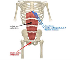
(1) Rectus abdominis
Origin: pubic crest and symphysis.
Insertion: costal cartilages of 5th, 6th, and 7th ribs, and xiphoid process of sternum.
Direction of fibers: vertical
Action:flexes the vertebral column by approximating the thorax and pelvis anteriorly. With
the pelvis flexed, the thorax will move toward the pelvis; with the thorax fixed, the pelvis
will move toward the thorax.
Weakness: a weakness of this muscle results in a decrease in the ability to flex the
vertebral column. In the supine position, the ability to tilt the pelvis posteriorly or to
approximate the thorax toward the pelvis is decreased, making it difficult to raise the head
and upper trunk. In order for anterior neck flexors to raise the head from a supine position,
it is essential that anterior abdominal muscles, particularly the rectus abdominis, fix the
thorax. With marked weakness of abdominal muscles an individual may not be able to
raise the head even though neck flexors are strong. In the erect position, weakness of this
muscle permits an anterior pelvic tilt and a lordotic posture (increase anterior convexity of
the lumbar spine).
(2) External oblique, anterior fibers
Origin: external surface of rib five through eight interdigitating with serratus anterior.
Insertion: into a board, flat aponeurosis, terminating in the lines alba, a tendinous raphe
which extends from the xiphoid.
Direction of fibers: the fibers extend obliquely downward and medialward with the
uppermost fibers more medialward.
Action: acting bilaterally, the anterior fibers flex the vertebral column approximating the
thorax and pelvis anteriorly, support and compress the abdominal viscera, depress the
thorax, and assist in respiration. Acting unilaterally with the anterior fibers of the internal
oblique on the opposite side, the anterior fibers of the external oblique rotate the vertebral
column, bringing the thorax forward (when the pelvis is fixed), or the pelvis backward
(when the pelvis is fixed). For example, with the pelvis fixed, the right external oblique
rotates the thorax counterclockwise, and the left external oblique rotates the thorax
clockwise.
 |
| External abdominal oblique (Ref: https://learnmuscles.com/) |
(3) External oblique, lateral fibers
surfaces of 10th, 11th and 12th ribs, interdigitating with latissimus dorsi.
Insertion: as the inguinal ligament, into anterior superior spine and pubic tubercle, and into
the external lip of anterior one half of iliac crest.
Direction of fibers: fibers extend obliquely downward and medialward, more downward than the anterior fibers.
Action: acting bilaterally, the lateral fibers of the external oblique flex the vertebral column,
 |
| Surface anatomy of external abdominal oblique (Ref: https://biologydictionary.net/) |
(4) Internal oblique, lower anterior fibers
Origin: lateral two thirds of inguinal ligament, and shirt attachment on iliac crest near anterior
superior spine.
Insertion: with transversus abdominis into crest of pubis, medial part of pectineal line, and into linea
alba by means of an aponeurosis.
Direction of fibers: fibers extend transversely across lower abdominal.
Action: the lower anterior fibers compress and support the lower abdominal viscera in conjunction
with the transversus abdominis.
(5) Internal oblique, upper anterior fibers
Origin: anterior one thirds of intermediate line of iliac crest.
Insertion: linea alba by means of aponeurosis.
Direction of fibers: fibers extend obliquely medialward and upward.
Action: acting bilaterally, the upper anterior fibers the vertebral column, approximating the
thorax and pelvis anteriorly, support and compress the abdominal viscera, depress the
thorax, and assist in respiration. Acting unilaterally, in conjunction with the anterior fibers
of the external oblique on the opposite side, the upper anterior fibers of the internal oblique
rotate the vertebral column, bringing the thorax backward (when the pelvis is fixed), or
the pelvis forward (when the thorax is fixed). For example, the right internal oblique rotates
the thorax clockwise, and the left internal oblique rotates the thorax counterclockwise on
a fixed pelvis.
 |
| Internal abdominal oblique muscle (Ref: https://learnmuscles.com/) |
(6) Internal oblique, lateral fibers
Origin: middle one thirds of intermediate line of iliac crest, and thoracolumbar fascia.
Insertion: inferior borders of 10th, 11th, and 12th ribs and linea alba by means of
aponeurosis.
Direction of fibers: fibers extend obliquely upward and medialward, more upward than the anterior fibers.
Action: acting bilaterally, the lateral fibers flex the vertebral column, approximating the
thorax and pelvis anteriorly, and depress the thorax. Acting unilaterally with the lateral
fibers of the external oblique on the same side, these fibers of the internal oblique laterally
flex the vertebral column, approximating the thorax and pelvis. These fibers also act with
the external oblique on the opposite side to rotate the vertebral column.
(7) Transversus abdominis (TrA)
Origin: inner surfaces of cartilages of lower six ribs, interdigitating with the diaphragm;
thoracolumbar fascia; anterior three fourths of internal lip of iliac crest; and lateral one third
of inguinal ligament.
Insertion: linea alba by means of a board aponeurosis, pubic crest and pecten pubis.
Direction of fibers: transverse
Action: acts like a girdle to flatten the abdominal wall and compress the abdominal viscera;
the upper portion helps to decrease the infrasternal angle of the ribs as in expiration. This
muscle has no action in lateral trunk flexion except that it acts to compress the viscera and
stabilize the linea alba, thereby permitting better action by anterior trunk muscles.
Weakness: permits a bulging of the anterior abdominal wall, thereby indirectly tending to
affect an increase in lordosis. During flexion in the supine position, and hyperextension of
the trunk in the prone position, there tends to be a bulging laterally if the transversus
abdominis is weak.
 |
| Transversus abdominis muscle (Ref: https://learnmuscles.com/) |
Transversus abdominis has been of particular interest to many physiotherapists as
a core stability muscle due to its anatomy. The influence of lumbar stability on poor posture
versus upright posture has also been studied. It has been reported that there is a significant
decrease in activity of the internal oblique and multifidus muscles in poor sitting and
standing postures.
The present study was an investigation into the changes in TrA thickness in commonly
adopted poor postures (sway-back standing and slouched sitting) compared to equivalent
neutral spine postures. The results show a significant thickening of TrA in both lumbo-pelvic
neutral erect standing and sitting postures compared to sway-back standing and slouched
sitting. TrA thickness has been shown to be correlated with muscular activity. Therefore,
the observed increase in thickness of TrA in erect standing compared to the sway-backed
position suggests that there is more TrA activity in erect standing. This increase in activity
may help to stabilize the spine.
For healthy abdominal muscles, we need to maintain good standing and sitting
posture, stretching, and strengthening.
Reference:
https://www.researchgate.net/publication/24428336_Effects_of_posture_on_the_thickness_
of_transversus_abdominis_in_pain-free_subjects
Kendall FP., et al. Muscles testing and function. Fourth edition. Williams & Wiikins. USA.
1993.


