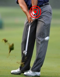
 |
| Ref: https://fittergolfers.com/ |
Wrist hyperextension
injuries do not develop only ligament or joint instability. It is an occasion
to develop pathology on tendon, bone, nerve, and vascular, as well. The biggest
cause is overuse induced degenerative that
is the result from sports activities
and daily activities such as occupational.
Extensor Pollicis Longus (EPL) Tenosynovitis
This
is one of tendinopathy and tendon instability. It is described as a drummer’s
palsy in which stenosing tenosynovitis of the extensor pollicis longus (EPL) is
seen in patients subject to long-term repetitive wrist hyperextension. The most
common has been seen in gymnasts and platform divers that the pathomechanics
are thought to involve impingement of the EPL tendon between the base of the
third metacarpal and the Lister tubercle, leading to inflammation, swelling,
and a subsequent discrepancy in size between the EPL and its tight, inelastic
fibrous compartment.
 |
| Ref: https://vectormine.com/ |
Tendon gliding limitation through the third
compartment affect a painful snapping sensation and can progress to attenuation
and rupture of the tendon. Traumatic injuries in this area can disrupt the
tendon’s blood supply or cause compressive swelling (e.g., hematoma formation)
within the third extensor compartment which leads to ischemic injury.
 |
| Platform diver (Ref: https://www.pinterest.co.uk/) |
Patients
are evaluated with pain and swelling around the Lister tubercle. Palpable
clicking or snapping may be felt with EPL firing in cases of stenosing
tenosynovitis. Radiographs and MRI can be useful in identifying any bony prominence
as a source of attritional tendon injury associated with synovitis or a
fracture in the setting of a recent trauma. Sonography is a helpful imaging tool
to investigate tendinosis and tenosynovitis, as well.
 |
| Sonography or ultrasound image (Ref: http://highlandultrasound.com/) |
Surgical
is generally recommended to treat. While corticosteroid injections may provide
a period of pain relief. However, these are typically avoided in athletes
because they can lead to tendon attenuation and increased risk for
rupture.
Extensor Carpi Radialis Brevis (ECRB) Insertional
Tendinitis
In high - level athletes
such as gymnastics, weight lifting, and racquet or stick sports (eg, baseball,
tennis, golf) bring repetitive forceful contraction that can cause microtrauma
to the tendinous insertion of the ECRB. Long-standing tenosynovitis eventually
leads to interstitial tendinosis and tendon attenuation.
 |
| Wrist extension during clean & jerk weight lifting (Ref: https://www.gymreapers.com/blogs/) |
It has been seen in
individuals, for example, construction workers and secretaries, who perform
repeated resisted forearm rotation, wrist extension, and prehension activities.
Activity-related pain
over the base of the second and third metacarpals should be done in typical
evaluation. In golf, baseball, and lacrosse athletes, the pain is typically in
the dominant hand and reproduced at the top of the backswing maximal wrist
extension; pain can also occur at the point of impact with the ball (e.g.,
golf, baseball). On physical examination, there may be point tenderness,
swelling, and bogginess over the base of the third metacarpal. Pain with
resisted wrist extension and passive wrist flexion is suggestive of ECRB
insertional tendinitis. MRI scans will show edematous changes to the distal
ECRB and its insertion.
 |
| The top of the backswing maximal wrist extension in golfer (Ref: https://www.bunkered.co.uk/) |
Conservative treatment
is the first consideration in the early phase primarily via rest/activity
avoidance and use of NSAIDs. Corticosteroid injections can be helpful to reduce
inflammation and pain but under caution, as they may lead to tendon attenuation
and risk for rupture. Goals for nonoperative treatment need physiotherapy to
complete symptom relief and full range of motion by 6 weeks, followed by 2
weeks of gradual strengthening and initiation of sport-specific training around
week 12, after the patient’s wrist has reached 85% of the strength of the
contralateral side.
 |
| Lacrosse (Ref: https://cmsvathletics.com/) |
Patients with mild
symptoms or faster progression through this general protocol may return to
sports sooner. Tenosynovectomy, the surgery, is indicated after 6 to 12 months
of failed nonoperative treatment. Postoperative, patients have their wrists
immobilized for 2 weeks, followed by a range of motion therapy. At 6 weeks
postoperative, the rehabilitation protocol is the same as the nonoperative
treatment described above.
Fourth-Compartment Syndrome
The extensor indicis
proprius (EIP) muscle originates along the distal third of the ulna and passes
within the fourth compartment, deep and ulnar to the extensor digitorum
communis (EDC) tendons.
 |
| Extensor Indicis Proprius (Ref: https://proper-cooking.info/) |
Increasing of the space
occupied within the fourth compartment was known in term “Anomalous” that can
cause pathologic increase in compartment pressure with subsequent
tenosynovitis, irritation of the posterior interosseous nerve (PIN), pain, and
disability. This is pathomechanics to develop forth – compartment syndrome.
Initial treatment is
typically nonoperative, with rest, NSAIDs, activity modification, splinting,
and corticosteroid injections. Patients who do not respond to prolonged
nonoperative treatment should raise suspicion for the presence of aberrant
anatomy (e.g., anomalous muscle or tendon). Surgery is indicated for patients
without improvement despite 3 to 6 months of nonoperative treatment, which
involves decompression via surgical release of the fourth extensor compartment;
concomitant tenosynovectomy and reduction or excision of associated anomalous
muscles may be performed to decrease the risk of recurrence, particularly in
patients who plan to return to sports.
Distal posterior interosseous nerve (PIN)
Syndrome
The PIN is the terminal
branch of the radial nerve, which passes through the 2 heads of the supinator
and travels to the wrist along the radial floor of the fourth extensor
compartment, just under the Lister tubercle. Terminal sensory branches of the
PIN cross dorsally over the scapholunate ligament and innervate the dorsal capsule
of the wrist.
 |
| Posterior Interosseous Nerve (PIN) (Ref: https://casereports.bmj.com/content/14/10/e245659) |
Athletes whose sports
require repetitive, forceful hyperextension of the wrist (e.g., gymnasts,
football linemen and defensive backs, platform divers, weight lifters),
particularly those with hypermobility at baseline, may experience dorsal wrist
pain secondary to impingement of the PIN at the wrist.
On examination will have
pain exacerbated by maximal dorsiflexion of the wrist as well as tenderness
localized to the fourth extensor compartment along the course of the PIN.
 |
| Gymnasts on balance bar (Ref: https://blog.orthoindy.com/) |
Initial treatment for
athletes with suspected distal PIN impingement is nonoperative by
immobilization and NSAIDs. Surgical treatment is indicated when PIN neurectomy
has been shown to be a safe and effective procedure for providing pain relief
in most patients.
Avascular Necrosis of the Lunate (Kienböck
Disease)
Sometimes, wrist pain is
caused by not enough or a blocked blood supply. Presenting symptoms are often
similar to those of wrist sprain without a history of trauma. Dorsal wrist
tenderness over the lunate with adjacent reactive synovitis and soft tissue
swelling is common. Decreased grip strength and pain with motion are usually
present and exacerbated by activity, particularly with extension and axial
loading across the wrist (e.g., push-ups or military press).
 |
| Military press or overhead press (Ref: https://www.inspireusafoundation.org/) |
Kienböck disease refers
to avascular necrosis of the lunate and is the most common type of idiopathic
carpal avascular necrosis. Its origin remains unclear and is likely
multifactorial, with local vascular and osseous abnormalities being most
commonly implicated. It is most common in men aged 20 to 40 years.
MRI and the presence of
uniform signal change of the lunate compared with the rest of the carpus are
used for diagnosis.
 |
| Avascular Necrosis of the Lunate (Kienböck Disease) MRI: Black area at carpal (Ref: https://www.orthobullets.com/) |
Patient symptoms and
radiographic staging of disease are the major treatment guidelines. Symptomatic
patients in early stages of disease are typically treated initially with cast
immobilization, in order to improve lunate vascularity. In later stages,
palliative and performed in an attempt to limit continued carpal collapse (e.g.,
proximal row carpectomy, wrist arthrodesis, denervation).
Occult Dorsal Carpal Ganglion
Occult dorsal ganglion
cysts may result from athletic activity and lead to a dorsal impingement
syndrome. 60%-70% of these mucin-filled cysts originate from the Scapholunate
ligament and most commonly present as a cystic mass extruding between the
extensor pollicis longus and extensor digitorum communis tendons.
Smaller, occult dorsal
wrist ganglions are more difficult to identify. An inciting injury to the SL
ligament and subsequent degenerative change is thought to lead to formation of
occult ganglion cysts, although an inciting injury is only reported in about
10% of patients.
 |
| Dorsal Carpal Ganglion (Ref: https://quizlet.com/) |
Diagnosis should be
considered for all athletes with dorsal wrist pain that becomes worse with
dorsiflexion and loading across the wrist joint. They will have maximal
tenderness over the SL interval, which is identified by palpating the soft
tissues directly over the inline of the Lister tubercle, which may be
exacerbated by passive hyperextension of the wrist.
Initial treatment
is nonoperative, with corticosteroid injection directly into the wrist capsule
followed by a period of splint immobilization, which can provide pain relief
and help with diagnosis. Surgical intervention is effective for patients with
significant activity-limiting pain and nonoperative treatment that has
failed.
Dorsal Capsular Impingement
Dorsal wrist impingement (DWI)
refers to a disorder characterized by mid dorsal wrist pain attributed to
capsulitis or synovitis of redundant capsular tissue impinging between the ECRB
tendon and dorsal ridge of the scaphoid. The onset may be relatively minor but
leads to swelling and thickening of capsular tissue that is prone to recurrent
episodes of impingement and a cycle of aggravation with persistent
inflammation.
 |
| Dorsal wrist impingement (Ref: https://journals.sagepub.com/doi/full/10.1177/23259671221088610) |
In chronic cases, osteophytes may
develop along the dorsal scaphoid, lunate, or dorsal rim of the distal radius,
which leads to worsening impingement and dorsal impaction. Pain is localized to
the ECRB, where it passes over the dorsal scaphoid, which is exacerbated with
full wrist extension and loading of the wrist in an extended position (e.g.,
tabletop push-off test).
Plain-film radiographs are
typically normal, and CT scans may show the development of small osteophytes.
MRI scans can be helpful in confirming DWI, which may show dorsal capsular
thickening and redundancy with signs of inflammation in this area.
.jpg) |
| ECRB anatomy (Ref: https://www.orthobullets.com/) |
The most DWI can be cured by
conservative within 2 to 3 months by rest, splint immobilization, and NSAIDs.
Corticosteroid injections are helpful to break the cycle of capsular
inflammation and swelling and often provide significant (70%) pain relief for
several weeks. Surgical treatment may be indicated for refractory cases that
fail nonoperative management.
Operative is needed If conservative
treatment cannot solve. Postoperatively, patients are placed in a removable
wrist orthosis and begin immediate range of motion therapy, with the goal of
full wrist motion at 2 to 3 weeks. Strengthening begins after full motion is
achieved, and athletes may begin a return-to-sports protocol around 6 weeks
postoperatively, when strength is 80% that of the contralateral side.
These most anatomy and
pathologies have talked about Gymnastic, Lister tubercle, second metacarpal and third
metacarpal. The treatment consists of conservative such as immobilize with
brace and strengthening therapeutic exercise, and surgery that need post
operative rehabilitation.
In fact, there are many wrist disorders syndrome in Sports or daily living that we will discuss together later.
Reference:


ไม่มีความคิดเห็น:
แสดงความคิดเห็น