วันศุกร์ที่ 13 พฤษภาคม พ.ศ. 2565

Physiotherapy with 11 ways to stretch the golfer's elbow.

Have you stretched your hand while typing? What pose do you stretch? I guessed it could be a forehand stretch which releases hand tension.  

Ref: https://www.avera.org/

11 ways to stretch the golfer's elbow. 

Exercise #1: Golfer’s elbow stretch with hand open: Direction to stretch the all five muscles is wrist extension with ulnar deviation with supination with straight fingers. 


Exercise #2: Golfer’s elbow stretch with hand closed: Direction to stretch Pronator teres and Flexor carpi radialis is wrist extension with ulnar deviation with supination


Exercise #3: Standard forehand supinate with hand open stretch


Exercise #4: Standard forehand supinate with hand closed stretch 


Exercise #5: Arm prayer stretch


Exercise #6: Standard forehand pronate with hand open stretch 


Exercise #7: Standard forehand pronate with hand closed stretch


Exercise #8: Table forehand pronate with hand open stretch


Exercise #9: Table forehand supinate with hand open stretch


Exercise #10: Floor forehand pronate with hand open stretch


Exercise #11: Floor forehand supinate with hand open stretch 


According to forehand muscle anatomy originates from the medial epicondyle of the humerus. From the radial to the ulnar aspects of the forearm, the musculature includes the pronator teres, the flexor carpi radialis, the palmaris longus, the flexor digitorum superficialis, and the flexor carpi ulnaris. The pronator teres and flexor carpi radialis both attach to the anterior aspect of the medial epicondyle. They are known as flexor - pronator muscles of the forearm.

The tendon of these five muscles concur at the common flexor tendon where is approximately 3 cm long and, in most elbows, crosses the ulnohumeral joint medially. 

Wrist flexor muscles anatomy
(Ref: https://www.flickr.com/photos/nickbrazel/136518076)


One of the elbow pain is epicondylitis. It is one of the most common elbow problems in adults that occurs both laterally and medially. The number of medial epicondylitis is much less frequently than lateral epicondylitis seven to ten times more often, approximately.

The majority of the disorder’s primary etiology is a repetitive stress or overuse of the flexor-pronator musculature. Excessive repetitive stress on the tendon eventually results in microtrauma and degeneration. Histopathologic examination has revealed a staged process of pathologic tendon change. Initially, repetitive trauma results in peritendinous inflammation. Continued injury results in angiofibroblastic hyperplasia, an invasion of vascular and fibroblastic elements into the tendon. Eventually, replacement of the normal tendon with angiofibroblastic hyperplasia results in structural breakdown and irreparable fibrosis or calcification. Activity causing such pathology to the common flexor tendon can eventually transfer forces deeper to the Ulnar Collateral Ligament, which mirrors Common Flexor Tendon fiber orientation and histologic anatomy.

 Degenerative changes in the musculotendonous region of the medial epicondyle are the result of chronic repetitive concentric and eccentric contractile loading of the flexor-pronator group. Most often such changes are seen in the pronator teres and the flexor carpi radialis muscles, although larger diffuse tears can occur in the palmaris longus, flexor digitorum superficialis, and flexor carpi ulnaris. Although repetitive overuse has been identified as the primary etiology, a single traumatic event, such as a direct blow or a sudden, extreme eccentric contraction, may result in the development of epicondylitis. Medial epicondylitis has been associated with activities involving repetitive forearm pronation and wrist flexion.

Ref: https://www.washingtonpost.com/


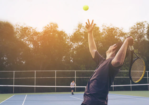
All activities, both sports and non - sports,  which require repetitive forearm, wrist, and hand motions. Sports activities; include,  golf, tennis, bowling, racquetball, football, archery, weightlifting, javelin throwing, and  baseball pitchers, resulting from intense valgus forces on the medial elbow during the late cocking and acceleration phases of throwing. For non - sports are  associated with occupations such as carpentry, plumbing, meat cutting, etc.

Ref: https://www.istockphoto.com/


The majority of the disorder’s primary etiology is a repetitive stress or overuse of the flexor-pronator musculature. Excessive repetitive stress on the tendon eventually results in microtrauma and degeneration. Degenerative changes in the musculotendonous region of the medial epicondyle are the result of chronic repetitive concentric and eccentric contractile loading of the flexor-pronator group. 

Activity causing such pathology to the common flexor tendon can eventually transfer forces deeper to the ulnar collateral ligament, which mirrors common flexor tendon fiber orientation and histologic anatomy. Most often such changes are seen in the pronator teres and the flexor carpi radialis muscles, although larger diffuse tears can occur in the palmaris longus, flexor digitorum superficialis, and flexor carpi ulnaris. Although repetitive overuse has been identified as the primary etiology, a single traumatic event, such as a direct blow or a sudden, extreme eccentric contraction, may result in the development of epicondylitis. 

 Histopathologic examination has revealed a staged process of pathologic tendon change. Initially, repetitive trauma results in peritendinous inflammation. Continued injury results in angiofibroblastic hyperplasia, an invasion of vascular and fibroblastic elements into the tendon. Eventually, replacement of the normal tendon with angiofibroblastic hyperplasia results in structural breakdown and irreparable fibrosis or calcification. Activity causing such pathology to the common flexor tendon can eventually transfer forces deeper to the ulnar collateral ligament, which mirrors common flexor tendon fiber orientation and histologic anatomy.

The inflammatory of musculotendinous origin at the medial epicondyle of the elbow is medial epicondylitis that is commonly referred to as “golfer’s elbow”. 

Ref: https://capitalchirodsm.com/tennis-elbow-versus-golf-elbow/


Although, stretching before performance may impact on some types of injuries but not impact on other injuries. Stretching benefits consist of increased flexibility, decrease pain, physiotherapy treatment process, injury prevention, basically. A plausible theory of injury prevention; include, (1) stretching makes the muscle–tendon unit more compliant, (2) increased compliance shifts the angle–torque relationship to allow greater relative force production at longer muscle length, and (3) subsequently the enhanced ability to resist excessive muscle elongation may decrease the susceptibility to a muscle strain injury.

 This theoretical rationale for why pre-participation muscle stretching might decrease the risk of subsequent muscle strain injuries is a testable hypothesis that has not been adequately addressed in the literature. Indeed, a counter hypothesis could be that enhanced contractile force production when a muscle is in a lengthened position could increase the likelihood of injury. Importantly, this rationale does not apply to the risk of other injuries such as ligament injuries, fractures or overuse injuries, such as tendinopathies.

Insertion of forearm flexor tendons in golfer’s elbow consist of wrist joint and fingers joint. Referring to anatomy, the way to stretch can be done with hand open and with hand closed. However, there are various poses to stretch the golfer’s elbow that are more advanced than hand closed. 

Conclusion, I would like to recommend stretching often to prevent injury because it affects the muscle - tendon unit which is the most common pathologic changed degeneration. The stretching principle is very simple: stretch to the point where “tightness without pain” or “noticeable tension without pain” will hold at the point for 30 seconds of 3 - 4 repetitions in one muscle as VIDEO demonstraion.


Reference:

https://journals.lww.com/jaaos/fulltext/2015/06000/medial_epicondylitis__evaluation_and_management.4.aspx


https://www.researchgate.net/publication/8474346_Stretching_and_injury_prevention_-_An_obscure_relationship/link/02bfe50f46bf7022d9000000/download


https://www.researchgate.net/publication/40766291_To_stretch_or_not_to_stretch_The_role_of_stretching_in_injury_prevention_and_performance/link/59ddfbc80f7e9bec3bae07b6/download 


วันอาทิตย์ที่ 8 พฤษภาคม พ.ศ. 2565

Physiotherapy with 3 shortcut exercises for forward head and neck pain.


            Forward head posture is one of the most common abnormal body characteristics which have been seen regularly. It is defined as front displacement of the head, together with over – backward bending of neck vertebral spine. This position shows round shoulders, increasing of anterior cervical convexity, and a decrease of craniovertebral angle which is the angle between the line from the external auditory meatus to the horizontal line at the level of the seventh cervical vertebra.

The forward head posture increases the loads on the necks and shoulders; it causes neck and back pain in 30% of monitor staff workers. Due to, the head is in front of the body-center that leads to a 3.6 times greater load than normal posture. Consequence is work related musculoskeletal disorders, such as pains of neck, shoulder, and headaches.

The forward head posture was first reported through investigation of muscle imbalance syndromes with a reduction in the length of the upper trapezius, the posterior part of the cervical extensor muscles (suboccipitals, semispinalis capitis, splenius capitis-cervicis), the sternocleidomastoid and the scapular elevator. Moreover, the Longus Capitus which is a deep neck flexor gets weak that causes Sternocleidomastoid fire first. As Sternocleidomastoid receives overactive tension, tone, and fatigues, this influences disability and neck pain in patients.  


To solve this problem needs to improve posture and reduction of tone and pain intensity. Therapeutic exercise protocols include general exercise and specific exercises aimed at strengthening particular muscles and soft tissue release technique.

             Strengthening of the Longus Capitus, the deep neck flexor, is a crucial specific strengthening exercise that does chin tuck as goal standard. This exercise can be done alone or combined with other muscles if necessary, for example, the latissimus dorsi pull down, shoulder external rotation exercise, prone horizontal shoulder abduction exercise, shoulder abduction exercise, shoulder flexion exercise and shoulder extension exercise. These exercises contribute shoulder and scapular stability that can improve rounded shoulders.  

             Stretching exercise of the upper trapezius, the posterior part of the cervical extensor muscles (suboccipitals, semispinalis capitis, splenius capitis and splenius cervicis), the sternocleidomastoid and the scapular elevator to release tension and increase specific range of motion in essential direction for reverse forward head.

             Some researchers said prolonged forward head posture will develop wrong alignment memory of neuromuscular. Therefore, neutral head position training to correct posture as a new habit is one of the important components.  


This article concluded 3 shortcut exercises to walk away from forward head and neck pain.

Exercise #1: Tennis ball massage at sub – skull


             Tennis ball or spike massage ball is between the sub skull and wall or floor. You can glide your head around and static compress for a while. Make this message until you feel release, then do other exercise


Exercise #2: Sternocleidomastoid stretch

             Start with bending the neck backward, then put both hands on the collar bone (clavicle), then slide both hands downward which holds 30 seconds for 5 reps, then start again. 


Exercise #3: Chin tuck exercise

             It sounds easy in terms of “glide your head backward” but some patients go in the wrong direction in the first session. The wrong direction includes bending the neck forward or bend neck backward so that you will see the floor or ceiling. Thus, the correct direction is to glide or slide the head backward so that you will look straight ahead. When gliding as far as possible, it is necessary to hold a few seconds and relax. According to exercise fundamentals are 15 reps/set for 3 sets. Intensive exercise needs only 10 reps for every hour during the day.


Remark: You can study various of neck stretching on https://rehabcompanion.blogspot.com/2022/04/physiotherapy-with-9-stretching_26.html


Reference:

https://www.researchgate.net/publication/344013528_A_Comparison_Study_of_Posture_and_Fatigue_of_Neck_According_to_Monitor_Types_Moving_and_Fixed_Monitor_by_Using_Flexion_Relaxation_Phenomenon_FRP_and_Craniovertebral_Angle_CVA


https://www.researchgate.net/publication/354683774_The_effects_of_therapeutic_exercise_in_improving_forward_head_posture_and_functionality_in_patients_with_neck_pain/link/61463e86519a1a381f6c1286/download


https://www.researchgate.net/publication/327397586_Efficacy_of_Suboccipital_and_Sternocleidomastoid_Release_Technique_in_Forward_Head_Posture_Patients_With_Neck_Pain_A_Randomized_Control_Trial/link/5cdd3766458515712eae3e2c/download


https://www.researchgate.net/publication/354683774_The_effects_of_therapeutic_exercise_in_improving_forward_head_posture_and_functionality_in_patients_with_neck_pain


วันจันทร์ที่ 2 พฤษภาคม พ.ศ. 2565

Physiotherapy with 8 rear shoulder structure stretching options


It is possible that the most common image of shoulder stretch must be “the cross body stretch” which has been online and offline. Shoulder Stretches are various poses and directions to involve front and rear muscles. Physiotherapists use shoulder stretches to contribute treatment with shoulder pain and shoulder health promotion. 

Rear shoulder structures stiffness or posterior shoulder tightness are related to many shoulder disorders and shoulder pain. They are muscles and shoulder capsule that can be impaired by disease such as frozen shoulder (shoulder capsulitis), rotator cuff injuries, rotator cuff repaired, etc or activities, such as, swimming, tennis, golf, baseball, volleyball, etc. 


“Posterior Shoulder Tightness has been defined in terms of reduced glenohumeral joint internal rotation and/ or glenohumeral horizontal adduction compared to the contralateral shoulder.”


Referring to this definition, I have seen more clinical cases that are able to develop limited shoulder elevation and glenohumeral head anterior translation. 

According to public articles, defining posterior shoulder tightness is considered a side-to-side difference ranging from 7 degrees to 20 degrees as an indication that posterior shoulder tightness is present. In the clinic, some of my patients with both shoulders internal rotation deficit, I compare to the normative range of motion. If shoulder internal rotation deficits greater than 10 % of normative, I will imply posterior shoulder tightness. By the way, the normative of shoulder internal rotation is 90 degrees. 

The relatively high incidence of posterior shoulder tightness in both athletic and nonathletic populations suggests its relevance to musculoskeletal shoulder pain. Posterior shoulder tightness is considered a contributor to posterior impingement, rotator cuff tendinopathy, subacromial impingement syndrome, collectively termed rotator cuff–related shoulder pain. Additionally, I have found in frozen shoulder (shoulder capsulitis) which is the stiffness of posteroinferior capsule and impaired rotator cuff muscle that accompany the posterior deltoid and triceps. 

This article demonstrates VIDEO to stretch rear shoulder structure for shoulder pain treatment in my clinic and can be patient instruction as a home program. 

Static stretching is the best way which I always suggest. Stretching to the point where “tightness without pain” or “noticeable tension without pain” will hold at that point for 30 seconds of 3 - 4 repetitions in each muscle.


8 rear shoulder structure stretching options 

Exercise #1: Sleeper stretch 


Exercise #2: Infraspinatus stretch 


Exercise #3: Cross body stretch


Exercise #4: Eagle pose stretch


Exercise #5: Traditional tricep stretch


Exercise #6: Modified triceps stretch


Exercise #7: Rhomboid stretch


Exercise #8: Shoulder pendulum: this is a special stretch of rotator cuff because it is a dynamic stretch which needs to draw clockwise and anti- clockwise circles 15 reps each direction for 3 - 5 sets. 


Reference: 

https://www.researchgate.net/publication/343293262_Posterior_shoulder_tightness_an_intersession_reliability_study_of_3_clinical_tests/link/5f298f6692851cd302dbe69f/download 


https://nismat.org/wp-content/plugins/zotpress/lib/request/request.dl.php?api_user_id=2488608&dlkey=VUJJ5786&content_type=application/pdf 


https://www.jospt.org/doi/pdf/10.2519/jospt.2007.2337


https://www.jospt.org/doi/pdf/10.2519/jospt.2018.0605 


วันอังคารที่ 26 เมษายน พ.ศ. 2565

Physiotherapy with 9 stretching exercise to improve rounded shoulders


Rounded shoulders
(Ref: Singla D., et al. Association Between Forward Head, Rounded Shoulders, and Increased Thoracic Kyphosis: A Review of the Literature. J Chiropr Med 2017;16:220-229)

            Rounded shoulder posture is one of the most common poor posture characteristics in physiotherapy clinics. In my experience, I see both cases of rounded shoulders accompanied with a forward head and only rounded shoulder alone. Chief complaint never talked about rounded shoulders; but, it is about muscle pain of neck, shoulder, upper back, and scapular. 

Rounded shoulder posture refers to a posture characterized by acromion protraction in front of the line of gravity, shoulder protraction, and downward rotation as well as anterior tilt.

    External cause factors, for example, computer and smartphone lifestyle, job characteristic, workstation, or temperature, ect. As internal cause factors reveal soft tissue impairment; include, weakness of scapular stabilizer muscles, upper back, rotator cuff, and shortening of chest muscle, upper arm muscle, some shoulder ligaments. 

In this physiotherapy article presents shortening of front upper part soft tissue shortening; and, how to stretch them.    


3 Major muscles involved rounded shoulders

            1. Pectoralis major

                The pectoralis major is a large, flat muscle of the pectoral girdle of the upper limb. It is a fan shaped that appears with 3 heads or portions: the clavicular, the sternocostal, and the abdominal head. This distinct tendon enters in contact towards the insertion on the humerus. Rounded shoulders is related to the shortening pectoralis major which pulls the humeral head anterior. It needs stretching and manual release from a physiotherapist to correct the posture.

Pectoralis major muscle
(Ref: https://link.springer.com/article/10.1007/s00064-021-00760-5)

            2. Pectoralis minor

                Pectoralis minor located between lateral 3rd, 4th, 5th rib to the coracoid process of scapular. When this muscle shortens, the scapular wiil be changed posture to be scapular anterior tilt, scapular internal rotation, and scapular downward rotation. It needs stretching and manual release from a physiotherapist as well.

Pectoralis minor muscle 
(Ref: https://www.orthobullets.com/anatomy/10007/pectoralis-minor)


            3. Biceps brachii

                Biceps brachii affects the shoulder joint becasue there are 2 different attachments of muscle; include, long and short heads. The long head of the biceps brachii is attached from the supraglenoid tubercle of the scapular and passes through the bicipital groove, and the short head of this muscle is attached from the coracoid process of the scapular.


Biceps brachii
(Ref: https://fitmachtgesund.de/en/musculus-biceps-brachii/)


The rounded shoulder posture as a result of lower trapezius muscle, serratus anterior muscle, and rhomboid muscle is prolonged lengthening that develops weakness and pain. In my experience, stretching and massaging them cannot improve pain. I always massage and stretch all 3 muscles which we talked about above. To exercise scapular stabilizer muscle is one of crucial treatments; but, it is not my point in this article. 

I prefer static stretching the most because it is more effective than dynamic stretching and easier than PNF stretching. The way to stretch is simple: stretch to the point where “tightness without pain” or “noticeable tension without pain” will hold at that point for 30 seconds of 3 - 4 repetitions in one muscle. 


Remark: If you feel neck and head discomfort from these stretching, you should decrease stretching range of motion. While these 3 muscles are very tight, the scapular and neck muscles try to beat them to achieve a range of motion that increases stress on scapular and neck muscles. Therefore, start at a small tension point and go further gradually.  


9 poses of stretching to improve rounded shoulders 


Exercise #1: Horizontal doorway pectoral stretch


Exercise #2: High doorway pectoral stretch


Exercise #3: Double shoulder external rotation stretch


Exercise #4: Active anterior deltoid stretch


Exercise #5: Chair anterior deltoid stretch


Exercise #6: Single horizontal pectoral stretch 


Exercise #7: Single shoulder external rotation stretch


Exercise #8: Half wall (chair) hang stretch


Exercise #9: Biceps brachii stretch


Reference:

https://he01.tci-thaijo.org/index.php/NurseNu/article/view/178634/143838 


https://www.researchgate.net/publication/318858178_Relationships_Between_Rounded_Shoulder_Posture_and_Biceps_Brachii_Muscle_Length_Elbow_Joint_Angle_Pectoralis_Muscle_Length_Humeral_Head_Anterior_Translation_and_Glenohumeral_Range_of_Motion/link/59a6a1ccaca272895c1671f2/download 


https://www.researchgate.net/publication/320543998_Changes_in_rounded_shoulder_posture_and_forward_head_posture_according_to_exercise_methods 


https://www.researchgate.net/publication/335136115_A_Detailed_Review_on_the_Clinical_Anatomy_of_the_Pectoralis_Major_Muscle/link/602e4fdb92851c4ed57c6f02/download 


Sports physiotherapy management for tennis elbow and other treatment options.

Ultrasound therapy in tennis elbow treatment (Ref: https://nesintherapy.com/) Tennis elbow is degeneration of the tendons that attach to t...