วันศุกร์ที่ 29 กรกฎาคม พ.ศ. 2565

Tips of proximal quadriceps stretch for strong stiffness with lower back pain

Ref: https://www.bicycling.com/


Quadriceps muscle is bi - articular joint muscle of hip and knee. Its function includes straight knee joint and flex hip joint that stretch the lower back, gluteal and hamstring. Normally, tightness of the quadriceps develops knee bending limitation that when bending the knee, the patient will feel tension at the muscle belly.


My physiotherapy experience, I have seen tightness of the proximal quadriceps in IT band syndrome, upper gluteal pain, and low back pain sometimes. If I take care of these cases, I will add on the proximal quadriceps assessment for more information. Mention to anatomy, this muscle can irritate pelvic posture due to the origin of attachment on a part of the pelvic where is AIIS. 

Quadriceps origin (red mark)
(Ref: https://compedgept.com/blog/)


Sometimes, I have found tightness of the proximal quadriceps following tightness of TFL. Perhaps, their origins are very near and they are located like a neighborhood. Sometimes, I have found only one of them gets tight. However, I would like to recommend stretching the proximal quadriceps if it demonstrated tightness. It can help to release rear side pain and improve posture. 

Strong stiffness of proximal quadriceps that cannot straight hip joint from flexion position.


Previously, I presented the way to stretch quadriceps. I have seen some patients had very strong stiffness of that tissue that would be the threat of recovery. Some of them have done the stretching difficulty. The patients alway compensate i.e. arch lower back or cannot upright hip and torso. 

Arching at lower back to compensate


I found one tips of proximal quadriceps stretch for strong stiffness as this VIDEO

Exercise #1: Half kneeling with toe stand stretch: the target leg is on the knee with set ankle at neutral. Lean back and pelvic backward slightly without arching the lower back. We need hip joint to be neutral or extension.


Case sample 1

Case triathlon athlete who has got both lateral groin pain after cycling training. The patient denied low back pain and gluteal pain. The muscle length assessment found tightness of both proximal quadriceps and slight tightness of TFL. This case did not has gluteus medius weakness that did not persuade me to think of IT band syndrome. One of my treatment processes was isometric contraction of quadriceps before proximal quadriceps stretching as demonstrated VIDEO. 

Cycling posture demonstrates prolonged hip flexion with prolonged gluteal and lower back stretch
(Ref: https://www.giant-bicycles.com/)


Case sample 2

Case swimmer who has got one side of the upper gluteal pain that was worse pain by crawl stroke and butterfly stroke. The patient did not has low back pain and knee pain. Gluteal muscles got pain from pressing and weakness which was gluteus maximus. The gluteus medius was a normal strength that  did not persuade me to think of IT band syndrome. QL and back extensor muscle were not spasms. I almost concluded only inflammation of gluteus maximus muscle, but I have seen slight hip flexion in supine lying. Additionally, the quadriceps muscle mass looked massive that illustrated the groove between ASIS and quadriceps belly.

Middle posture demonstrate quadriceps look like massive and groove between ASIS and quadriceps. Right posture demonstrate neutral posture that not demonstrate groove between ASIS and quadriceps.
(Ref: https://www.kateskinnerpt.com/posture-and-positioning) 

            This groove was only on the pain side and not on the other one. I did more evaluation for quadriceps muscle length, then it showed tightness of the proximal quadriceps. One of my treatment processes was stretching proximal quadriceps and gluteus maximus facilitation. I gave a home program assignment for stretching as demonstrated VIDEO and exercise gluteus maximus. 


Case sample 3

Case of a computer office worker who has got one side of upper gluteal pain and neck pain with radiation to the lateral thigh and tibia from forward reaching to put something on the shelf 2 months ago. The patient was treated by medicine, physiotherapy modalities, and stretching of the gluteus and hamstring. Firstly, I considered about piriformis syndrome. The job characteristic is prolonged sitting at the working desk. The standing posture showed a torso shift forward. There was severe pain and hypersensitivity that felt pain at the gluteus maximus, gluteus medius, TFL, and quadriceps. Torso forward bending was limited by pain with a very narrow range. Torso backward bending was limited by worse pain and radiated to the lateral foot. It was not only pain but also numbness on some range of motion that made me think about nerve irritation.

One sample of stand and reach function in normal life living that can stress to lower back and gluteal
(Ref: https://depositphotos.com/)


            I started treatment with a gentle massage on my iliopsoas and quadriceps because firstly my aim was improve posture. It was a good response that pain intensity was decreased significantly. Then I started stretching iliopsoas and proximal quadriceps gradually where pain and numbness free. After the hip extension range increased, I started gluteus maximus facilitation to the hip stability function. Finally, the pain intensity decreased 70 - 80% with improved standing posture. The torso range of motion and all tenderness of gluteal muscle were improved. I gave a home program assignment for stretching as demonstrated VIDEO and exercise gluteus maximus. Moreover, the patients was recommended not to be prolonged sitting because proximal quadriceps may be tight together with prolonged stretch gluteal region. I concluded this case was upper gluteal strain with proximal quadriceps spasm.  

Prone hip extension exercise for strengthen gluteus maximus
(Ref: https://www.saintlukeskc.org/)


The principle to stretch this muscle is the same as the others: stretch to the point where “tightness with pain” or “noticeable tension without pain” will hold at the point for 30 seconds of 3 - 5 reputations following a demonstrated VIDEO. 


วันอาทิตย์ที่ 24 กรกฎาคม พ.ศ. 2565

Physiotherapy with 3 options to stretch latissimus dorsi for shoulder pain or back pain.


Ref: https://jackhanrahanfitness.com/


Anatomically, Latissimus dorsi is a large flat muscle attached between the half lower spine and shoulder. It plays a vital role in pathomechanics of mechanical low back pain. Including shoulder pain, upper back pain, and maybe neck pain because it can make scapular motion changes or scapular dyskinesis. 

Ref: https://www.quora.com/


In my physiotherapy experience, weakness and stiffness of latissimus dorsi develops poor posture. For example, I have named The Gorilla posture which I have seen in muscular men because of the stiffness of this muscle, moreover, I have called it as The swimmers posture which I have seen in swimmers athletes. It frequently causes shoulder pain in this posture. According to some reports, they mentioned to canoeists and rugby players that I never treated them. 

Ref: https://www.dreamstime.com/


Occasionally, I took care of a neck pain patient with breast augmentation. They had neck pain, upper back pain, or shoulder pain that could trick me to treat other muscles i.e. rotator cuff. The involved soft tissue of breast augmentation may result from incision scar and myofascial fascia stiffness and latissimus dorsi stiffness. 

Therefore, latissimus dorsi stretching is necessary for shoulder pain and back pain. 

3 poses option to stretch latissimus dorsi

Exercise #1: Stand latissimus dorsi stretch: to bend the torso to the opposite side of the target muscle that needs to place the hand on the wall for balancing and place the same foot behind the non - target side. Shift body weight to the hip which has the same target muscle side.


Exercise #2: Child pose lateral bending: start with child pose and bend the torso to the opposite side of target muscle.


Exercise #3: Gym ball latissimus dorsi stretch: this is floor exercise with the gym ball. Put non - target muscle on the ball, then place both feet anywhere which is the most stable. Elevate arm with close to the ear. 


The latissimus dorsi is primarily considered a muscle with actions at the shoulder, despite its widespread attachments at the spine. There is some dispute regarding the potential contribution of this muscle to lumbar spine function. 

Latissimus dorsi is a unique muscle in terms of its anatomical design. It is a relatively thin, fan-shaped muscle, and is one of the largest in the body in surface area. It covers a significant portion of the back to shoulder that there is some dispute regarding the potential contribution of this muscle to lumbar spine and shoulder function. 

Latissimus dorsi muscle (Bright red)
(Ref: https://www.physio-pedia.com/)


  Its attachments from the spinous processes of the lower six thoracic vertebrae, lumbar vertebrae, and sacrum, as well as to the ilium via the thoracolumbar fascia. Muscle fibers then converge superiolaterally toward the axilla via inferior 3 or 4 ribs and inferior angle of scapula , where the fibers converge to a single twisted tendon that is inserted in the intertubercular groove of the humerus which is onto the anterior aspect of the proximal humerus. 

Insertion of latissimus dorsi
(Ref: https://www.medicinebau.com/)


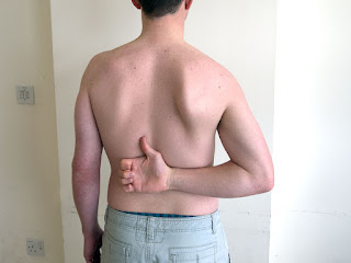
Muscle function of latissimus dorsi includes shoulder adducts, shoulder extends and shoulder internal rotates that are hand behind back. It provides a powerful rotator of the trunk and assist back extension. It is the antagonist of deltoid and trapezius muscle. The structural characteristics of this muscle are those of a global mobiliser. The muscle will therefore shorten, this will cause limited glenohumeral joint flexion and external rotation.

Hand behind back
(Ref: https://www.medistudents.com/)


The latissimus dorsi plays an important role in basic back activities such as trunk extension and rotation. There is a prevalence rate of 73% for latissimus dorsi tightness in active mechanical low back pain patients. A decrease in length or increase in stiffness of latissimus dorsi can lead to alterations in movement patterns and postures which cause exacerbation of low back pain that is a pathomechanics low back pain. A decrease in length or an increase in stiffness of this muscle can lead to alterations in movement patterns and/or postures which can cause an increase in low back pain. Latissimus dorsi in association with thoracolumbar fascia helps in force distribution that the posterior layer of thoracolumbar fascia which is formed through latissimus dorsi can effectively contribute to low back pain. 

Latissimus dorsi length test: normal length (left), stiffness (right)
(Ref: https://www.mitchmedical.us/)


The length of latissimus dorsi can be assessed by means of a reliable test to maintain treatment efficacy and objectivity. An latissimus dorsi length test explained by McConnell (1994) described a patient in crook lying. The patient has to decrease the lumbar lordosis actively by controlling the anterior pelvic tilt (by actively keeping the back flat) to do flexion with the glenohumeral joint in a neutral position with no glenohumeral rotation being allowed. The range of motion of glenohumeral joint flexion is measured using a goniometer at the point when the patient’s lumbar spine starts tilting anteriorly and lifts off the plinth, or when the glenohumeral starts to internally rotate. 


Discussion from above, to reverse muscle function for stretching this muscle should illustrate torso rotation and shoulder abduction. VIDEOs did not demonstrate torso rotation and shoulder abduction, however, torso lateral bending and shoulder adduction in flexion. Other motions are the same as above including shoulder flexion, shoulder external rotation, and lower back flexion.   

Ref: https://www.enjoy-swimming.com/


The principle to stretch this muscle is the same as the others: stretch to the point where “tightness with pain” or “noticeable tension without pain” will hold at the point for 30 seconds of 3 - 5 reputations following demonstrated VIDEO. 


Reference: 

https://www.scielo.cl/pdf/ijmorphol/v24n4/art30.pdf 


https://onlinelibrary.wiley.com/doi/full/10.1111/joa.12074 


https://www.sciencedirect.com/science/article/pii/S2095254613000215 


https://www.ijsar.in/Admin/pdf/to-study-the-prevalence-of-latissimus-dorsi-tightness-in-patients-with-mechanical-low-back-ache.pdf 


https://www.ijsr.net/archive/v9i8/SR20730124217.pdf 


https://www.researchgate.net/publication/323702778_Inter-_and_intra-rater_reliability_of_a_technique_assessing_the_length_of_the_Latissimus_Dorsi_muscle 


วันอังคารที่ 19 กรกฎาคม พ.ศ. 2565

Physiotherapy with 6 ways for QL stretching in non - specific low back pain


Ref: https://www.miamivalleygolf.org/

    Low back pain is usually classified as ‘specific’ or ‘non-specific’. Quadratus lumborum (QL) is one of the common sources of pain that can be overlooked easily. Specific low back pain is defined as that caused by a specific pathophysiological mechanism, such as disc prolapse or herniated nucleus pulposus, infection, inflammatory arthopathy, tumour, osteoporosis or fracture. 

Non - specific low back pain is defined as low back pain not attributable to a recognizable, known specific pathology. Quadratus lumborum pain syndrome is a myofascial pain syndrome. QL has a strong relationship with low back pain because of anatomy. The QL is located between the posterior pelvic and lumbar spine and 12th rib, which is an intimate lower back muscle. The pain is due to spasm and stiffness of the muscle. Many times, weak back muscles are compensated by quadratus lumborum leading to painful spasms.

Ref: https://radiopaedia.org/

In my physiotherapy experience, I have seen QL spasm and stiffness in patients with non - specific low back pain. They illustrated limited torso lateral flexion and tenderness of QL, additionally it may or may not cause spasm of the lower back muscle. This muscles always involve scoliosis condition. In the case of disc herniation, the QL muscle spasm never showed up. Patients with scoliosis have always seen the QL impairment that has been related to gluteus medius weakness. 

Neutral spine (left), Scoliosis spine (right)
(Ref: https://www.pinterest.com/)

6  ways to stretch QL 

Exercise #1: Basic stand torso lateral bending: Bend torso to opposite side of target muscle. Be careful: focus at torso, not head and neck.


Exercise #2: Wall torso lateral bending: Target muscle is next to the wall. Then, put the forearm and pelvic on the wall which is the target side. Remark: the case of lumbar disc herniation should stretch under supervision of physiotherapist. Additional, If you feel worse pain and/or leg's nerve irritation, should stop and consult physiotherapist.


Exercise #3: Side lying push up: Lying on the target muscle. Then, push up or on elbow for upper torso elevation with keep pelvic on the floor.


Exercise #4: Side lying on foam roll: Lying on foam roll or towel that put the non-target on foam roll.


Exercise #5: Cross sitting with lateral torso bending: Bend torso to opposite side of target muscle with hand support for more relax. Be careful: focus at torso, not head and neck.


Exercise #6: Half cross sitting with lateral torso bending: One leg is in front and other is rear leg. Bend torso to opposite side of target muscle that mean bend away from the front leg. Be careful: focus at torso, not head and neck.


Quadratus lumborum (QL) is a posterior abdominal wall muscle and bottom - up layout muscle.The QL originates from the posteromedial iliac crest and inserts into the medial border of the 12th rib and the transverse processes of the 1st to 4th lumbar vertebrae. The lateral free border of quadratus lumborum is angled from craniomedial to caudolateral.

Ref: https://www.physio-pedia.com/

    About their osseous attachments, the principal types of fascicles were iliocostal, iliolumbar, iliothoracic, and lumbocostal. These fascicles arose from the iliac crest passed to the 12th rib (iliocostal), from the transverse processes of lumbar vertebrae (iliolumbar), from the lateral surface of the 12th thoracic vertebra (iliothoracic), or from the lumbar transverse processes and passed to the 12th rib (lumbocostal) respectively. On the iliac crest the muscle occupied an area extending from 5 to 7 cm laterally from a point opposite the tip of the L4 transverse process. Occasionally, fascicles arose from the iliolumbar ligament instead of the iliac crest. On the 12th rib, fascicles attached to an area on the lower anterior surface that extended to between 4.5 and 7 cm from the head of the rib. 


It involves the muscle nearby including the quadratus lumborum and psoas major muscles traverse posterior to the lateral and medial arcuate ligaments of the diaphragm, respectively. The QL muscle lies in front of the erector spinae muscle group, consisting of the multifidus, longissimus, and iliocostalis. Moreover, one fascia is a fibrous composite of aponeurotic and fascial tissue that surrounds the QL which is the thoracolumbar fascia. 

QL is next to psoas, and both of them connect to diaphragm
(Ref: https://pocketdentistry.com/)

    The thoracolumbar fascia is part of a myofascial girdle that surrounds the lower torso and is important for posture, load transfer, and stabilization of the lumbar spine. It comprises multilayered fascia and aponeuroses, with two proposed models including the two-layered model and the three - layered model. Although the three-layered model is the most commonly used, regardless of which model is accepted. Because the fascial planes in the abdominal compartment follow the quadratus lumborum and psoas muscles through the medial and lateral arcuate ligaments and the aortic hiatus of the diaphragm, forming the endothoracic fascia.

In the three-layered model describes the posterior thoracolumbar fascia layer surrounds the erector spinae muscles, the middle layer passes between the erector spinae muscles and quadratus lumborum, and the anterior layer lies anterior to both quadratus lumborum and psoas muscles. 

The three-layered model of thoracolumpar fascia 
Ref: https://www.lecturio.com/


The characteristic of each layer was explained as below,

(1) The posterior layer: consisting of iliocostal fascicles laterally and iliolumbar fascicles medially. The iliocostal fascicles arose from the lateral third of the iliac attachment site but passed to all thirds of the costal attachment site, inserting behind or lateral the fascicles of the middle layer. The more lateral fascicles of the posterior layer typically became tendinous and appeared to insert into the middle layer of thoracolumbar fascia, but their tendons could be traced, through the fascia, into the 12th rib. The iliolumbar fibers arose from all thirds of the iliac site and passed to the tips of the upper lumbar transverse processes, most consistently to L3 and L1, and less often to L2 and L4. The posterior layer was wedge shaped in a longitudinal profile: thick inferiorly and tapering superiorly. This feature arose because, from below upwards, individual fascicles twisted, such that more lateral fibers covered their more medial companions, as the fascicle flattened out towards its costal attachment. As a result, fascicles with a thick, but narrow, origin from the iliac crest assumed a wide, but thin, linear insertion on the 12th rib.

(2) The intermediate layer: consisting of a set of lumbocostal fascicles. This layer was distinguished by the radiate arrangement of its fascicles. Most consistently, fascicles radiate from the tip of the L3 transverse process to various points on the costal attachment site of the muscle, behind the fascicles of the anterior layer. These fascicles were supplemented by similar fascicles arising most often from the L4 transverse process, and less often from the L2 and L5 transverse processes.

(3) The anterior layer: formed the anterior surface of the muscle, and presented a smooth quadrangular surface of regularly arranged fibers. It was a thin layer, consisting of iliocostal and iliothoracic fascicles, which assumed muscular or tendinous attachments at either end. The iliocostal fascicles arose from various points along the anterior margin of the attachment site on the iliac crest; only occasionally did fascicles arise from the iliolumbar ligament. They were distributed to various points along the entire attachment site on the 12th rib. The iliothoracic fibers arise from various points along the iliac site and converge to the lateral surface of the body of the twelfth thoracic vertebra. In some specimens they were joined by fascicles from the L4 and L5 transverse processes. In one specimen, a fascicle from the iliolumbar ligament reached the L1 transverse process.

Thoracolumbar fascia (green)
(Ref: https://bodyworksprime.com/)

The functions of the quadratus lumborum muscle are questionable. The QL clearly causes lateral flexion, raises the pelvic crest and is an important stabilizer of the lumbar spine. But uncertainty applies to extension that ‘probably helps to extend’ or ‘may extend’ the lumbar spine. 

A sustained contraction of QL is required during sitting, walking, lying, and other functional activities in order to stabilize the trunk and maintain body mechanics. Poor posture and body positioning alters the body mechanics which results in development of myofascial pain syndrome in this muscle. Therefore, treatment of QL disorders need not only stretching or massage, but also strengthening and postural control.  

Function of QL
(Ref: https://www.themedicalmassagelady.co.uk/)


    The principle to stretch this muscle is the same as the others: stretch to the point where “tightness with pain” or “noticeable tension without pain” will hold at the point for 30 seconds of 3 - 5 reputations following a demonstrated VIDEO. 


Reference:

https://www.researchgate.net/publication/330804178_Quadratus_Lumborum_Block_Anatomical_Concepts_Mechanisms_and_Techniques 


https://journals.sagepub.com/doi/pdf/10.1243/09544119JEIM266 


https://www.researchgate.net/publication/339043425_Effect_of_Stretching_on_Shortened_Quadratus_Lumborum_Muscle_in_Non_Specific_Low_Back_Pain


https://www.researchgate.net/publication/334834964_Atraumatic_Back_Pain_Due_to_Quadratus_Lumborum_Spasm_Treated_by_Physical_Therapy_with_Manual_Trigger_Point_Therapy_in_the_Emergency_Department 


https://www.semanticscholar.org/paper/Effectiveness-of-Strain-Counterstrain-Technique-on-Vohra-Jaiswal/ac6d022f234aab675aa87ef17657d513c7c9b383 


https://www.indianjpain.org/article.asp?issn=0970-5333;year=2018;volume=32;issue=3;spage=184;epage=186;aulast=Barge 


วันพฤหัสบดีที่ 14 กรกฎาคม พ.ศ. 2565

Physiotherapy with 7 options to stretch anterior shin muscle


Ref: https://www.sportsandspinal.net.au/)


            The muscles of the anterior compartment of the leg are listed in medio-lateral direction: tibialis anterior (TA), extensor hallucis longus (EHL), extensor digitorum longus (EDL), and peroneus tertius (PT). The name of them refer to “extensor” but they provide ankle dorsiflexion.

In my physiotherapy experience, they are difficult to feel stretched in the stretching procedure that I provide massage rather than stretching. However, stretching is necessary for them.

Heel to toe drop calculation
(Ref: https://www.permanent365.top/)


Sometimes, I have seen patients with anterior shin soreness after running. I asked them about how to run, type of running shoes, distance, changing speed as routine questions. The most was indicated to be tibialis anterior because of repetitive ankle dorsiflexion in running. By the way, running with low heel to toe drop will need more muscle workload for ankle dorsiflexion than high heel to toe drop. Distance and changing speed can be a risk factor if muscle fitness is not good enough. Because ground reaction force can impact every landing. 

Ankle inversion twisting
(Ref: https://www.marca.com/en/basketball/nba/)


In the case of ankle sprain with inversion twisting, I have always assessed extensor digitorum longus, peroneus tertius, peroneus longus, and peroneus brevis, especially if not any swelling at the ATFL ligament. The inversion direction can result in overstretching to their tendon, then develop muscle guarding for a protective mechanism. I have given it a stretch in proper period of healing. 


7 ways to stretch anterior leg compartment

Exercise #1: Seat tibialis anterior stretch: bend to plantarflexion with press foot downward.


Exercise #2: Seat peroneus tertius stretch: bend to plantarflexion with press foot upward.


Exercise #3: Seat EDL stretch: bend ankle with 4 toes (index toe to little toe) to plantarflexion.


Exercise #4: Seat EHL stretch: bend ankle with big toe to plantarflexion.


Exercise #5: Sit on heel stretch anterior ankle joint.


Exercise #6: Sit on heel to stretch EDL: sit on heel and 4 toes (index toe to little toe) are pulled to the plantar. 


Exercise #7: Sit on heel to stretch EHL: sit on heel and big toe is pulled to the plantar. 


(1) Tibialis anterior (TA)

The tibialis anterior muscle has a prismatic belly that arises from the lateral condyle of the tibia, proximal one-third to two-thirds of the lateral surface of the tibial shaft, anterior surface of the interosseous membrane, deep surface of the fascia cruris and intermuscular septum. The insertion of TA is a tendon that begins at about the level of the junction between the lower and middle thirds of the tibia and courses towards the medial border of the foot. The TA tendon inserting vertically on the first metatarsal base and the medial cuneiform bone. 

TA’s function for ankle dorsiflexion and ankle inversion. It also plays a role in suspension of the arch and controls supination of the rearfoot. Lesions of the tibialis anterior muscle and tendon are not frequently reported in international literature although pathology like tibialis anterior tendinosis or rupture is not rare.

Tibialis anterior muscle (green)
Ref: https://mobilephysiotherapyclinic.net/

(2) Extensor Hallucis Longus (EHL)

  The extensor hallucis longus is a thin muscle situated deep between the tibialis anterior muscle and the extensor digitorum longus (EDL). The EHL arises from the middle half of the fibula and from the interosseous membrane, medial to the origin of the EDL. The muscle belly becomes a long tendon that inserts through the tendon. Its tendon passes behind the superior and inferior extensor retinaculum, crosses the anterior tibial artery and vein from the lateral to the medial side near the ankle, and finally inserts on the dorsal aspect of the base of the distal phalanx of the big toe. 

The function of the EHL is to extend the big toe, dorsiflex the foot, adjunct foot eversion and inversion and stretch the plantar aponeurosis.  

Ref: http://npt.kr/fa/754

(3) Extensor Digitorum Longus (EDL)

Extensor Digitorum Longus has the topographical relationships on the lower leg that is on the anterior surface with the fascia of the leg and skin, medial to the tibialis anterior muscle and the extensor hallucis longus (EHL) muscle. 

EDL comes from the lateral condyle of the tibia, the proximal 2/3 of the anterior margin of the fibula, the superior part of the interosseous membrane, the deep fascia of the region and the anterior intermuscular septum.

Extensor Digitorum Longus (green)
(Ref: https://www.kenhub.com/en/)

It inserts through fibrous expansions of tendons 2nd - 5th onto the middle and distal phalanx of fingers 2nd - 5th. EDL has the following topographical relationships on the leg: the anterior surface with the fascia of the leg and skin; medially with the tibialis anterior muscle and the extensor hallucis longus muscle.

It produces extension of fingers 2nd - 5th and dorsal flexion of the foot, with an additional external rotation (pronation).


(4) Peroneus (Fibula) tertius (PT)

The Peroneus or Fibularis tertius muscles sound like it is in the lateral leg region, same as peroneus longus and brevis, but it is part of the anterior leg region.  

Peroneus tertius originates from the distal part of the fibula, the interosseous membrane, and anterior intermuscular septum as a derivation of the extensor digitorum longus muscle.  

Ref: https://stock.adobe.com/


Its inserting into the base of the fifth metatarsal, and also may be inserted in the shaft of the 5th metatarsal and  through a thin expansion on 4th interosseous space, the shaft of the fourth metatarsal, or at the base of the fourth metatarsal. It is often described as part of EDL.

 PT functions as a crucial contributor in dorsiflexion and eversion, postulated over the years as a stabilizer of the talocrural joint, avoiding forced investment and protecting the anterior talofibular ligament.


Peroneus tertius (Yellow arrow)
(Ref: https://www.scielo.cl/pdf/ijmorphol/v28n3/art16.pdf)


The principle to stretch this muscle is the same as the others: stretch to the point where “tightness with pain” or “noticeable tension without pain” will hold at the point for 30 seconds of 3 - 5 reputations following demonstrated VIDEO. 


Reference: 

https://www.orthopaedicmedicineonline.com/downloads/pdf/B9780702031458000909_web.pdf 


https://www.researchgate.net/publication/262781650_Ultrasound_of_tibialis_anterior_muscle_and_tendon_Anatomy_technique_of_examination_normal_and_pathologic_appearance 


https://www.researchgate.net/publication/51114950_A_variation_of_the_extensor_hallucis_longus_muscle_accessory_extensor_digiti_secundus_muscle 


https://sciendo.com/it/article/10.2478/jim-2021-0025 


https://bmcmusculoskeletdisord.biomedcentral.com/track/pdf/10.1186/s12891-019-2688-8.pdf 


https://www.scielo.cl/pdf/ijmorphol/v28n3/art16.pdf


Sports physiotherapy management for tennis elbow and other treatment options.

Ultrasound therapy in tennis elbow treatment (Ref: https://nesintherapy.com/) Tennis elbow is degeneration of the tendons that attach to t...